Flow Cytometry Analysis

ฝัง
- เผยแพร่เมื่อ 22 พ.ค. 2024
- Flow cytometry is a technique that lets us analyze both populations of cells as well as characteristics of individual cells. During this process, antibody-tagged cells are passed through the laser of a flow cytometer to measure the fluorescence from the antibody tag as well as other characteristics of the cells. Learn how scientists can use this kind of analysis to compare the cells from different people
This video is part of the Research Integrated Science Education (RISE) Program that is funded by SENS Research Foundation and Dalio Philanthropies. The program is designed to introduce high school students to aging research and laboratory concepts, such as experimental design and data interpretation, within core curriculum standards. To learn more about RISE and to access additional functionalities such as the transcript glossary function, visit www.sens.org/rise - วิทยาศาสตร์และเทคโนโลยี


Best Explanation of all videos in the Internet. Thanks
This is the awesome explained video in all other explained garbage... Kudos
Thank you for making the content so intuitive! Well done!
Thank you for taking about the prepping process. Massive help.
This was awesome. I was SO confused abut how flow Cytometry actually worked at its most basic. Thank you!
Great video!! I am so excited to show this to my laboratory students! Thank you!
Good work! very informative for a beginner to follow all the little steps involved in the FACS analysis.
Awesome, I am a statistician in the pharma industry. I have no background in flow cytometry, however, it is the nature of my job that I frequently face with CyTOF data and have to make sense of it. This tutorial is really helpful. Thank you so much!
WOW! This video is amazing. So clear, so well produced. Thank you so much!
This video is very easy to understand. Great work!!
SUPERB explanation of flow cytometry!! Thanks a bunch for the work!!
thank you!! this was explained so well and made it easy to understand.
Great video, but I have to point out an error, has at 8:30 the side scatter and the forward scatter are wrongly attributed. Has their names suggest, the forward scatter is directly in front of the laser beam, and the side scatter is in perpendicular plane to it
Thank you! I was about to say that
Wow, impressive depth and conveyance of the material.
This was very helpful, thanks! I'm applying for a job in a lab that does flow cytometry and had never heard of it before. This was a great first look
very easy to understand, thank you for the video and great explanation
finally i can get it!!! thank you for making this so easy to understand
Excellent explanation, thanks and well done!
very awesome and easy to catch. Thank you very much!
Best cytometry flow explanation, thank u
The best flow cytometry video i have ever seen 👍👍👍👍👍
Isma colours dyes ki smj ae ha ?
Yes , finally I understand what Flow Cytometry is . Thanks a lot !!!
Thank you for the wonderful explanation
Very clear video. Thank you.
Thank you for explaining so well!
awesome video! thanks!!
Great video!
Really enjoyed and understood this …. Thank you
Woaw, excellent explanation, now I understand better. Thank you a lot!
Great job! Thank you for such a simple, to the point video explaining the basis of flow cytometry! Kudos!! I must say, it was cute when you mentioned "You must be thinking if cells come with grocery stickers.."!!❤
Excellent!!!! I had no idea of flow cytrometry and now I can say I'm quite a bit less ignorant...
Such a helpful video. Thank you
謝謝 講得很清楚
Great teaching !
Great video
Thank you!
AMAZING AMAZING video! VERY well done
amazing video!
Very nice and well explained video
Thank you
excellent, well done
Thank you SO MUCH
Excellent 👍🏾 thanks 🙏
Thanks 4 this. I am new to research methods and I was looking for a topic to present
So helpful
good work
THANK YOU SO MUCH
thanks for this video.
thank you
Best video for introduction to flow cytometry. Thank you so much, ❤️
Thank you! Glad it helped! Let us know if you have any questions or suggestions for future content!
@@SENSRF can you please upload a video on how to analyse cytometry graphs and how the cell population look on the graph animated and then how it look in cytometer
bravo !
Hello @SENS Research Foundation From minute 7:30. Why are you marking the detector in front of light source(right behind the object) as side scatter and the one that is placed at 45° as forward scatter? Isnt it other way around?
Thanks for reply,
Arti
Many thanks for great explanation but
Can I have a question?
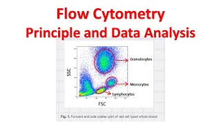
What does the colors of the dots indicate (as in 9:21). Is it the density? Like the number of cells in red area will higher than yellow one, and yellow > green > blue right?
sorry for my poor english
I didn't know how i thanks to you.
Why do cells that do not fluoresce (@11:51) show some small peaks? Why don't we have the peaks absent for such cells?
wow!
COOL
can you provide translation for arabic language? please
X5 speed