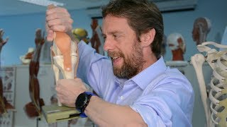
Knee Joint Anatomy Animation : Bones, Ligaments, Menisci, Innervation, Blood supply and Movements

ฝัง
- เผยแพร่เมื่อ 5 ต.ค. 2023
- 📌 𝐅𝐨𝐥𝐥𝐨𝐰 𝐨𝐧 𝐈𝐧𝐬𝐭𝐚𝐠𝐫𝐚𝐦:- / drgbhanuprakash
📌𝗝𝗼𝗶𝗻 𝗢𝘂𝗿 𝗧𝗲𝗹𝗲𝗴𝗿𝗮𝗺 𝗖𝗵𝗮𝗻𝗻𝗲𝗹 𝗛𝗲𝗿𝗲:- t.me/bhanuprakashdr
📌𝗦𝘂𝗯𝘀𝗰𝗿𝗶𝗯𝗲 𝗧𝗼 𝗠𝘆 𝗠𝗮𝗶𝗹𝗶𝗻𝗴 𝗟𝗶𝘀𝘁:- linktr.ee/DrGBhanuprakash
Knee Joint Anatomy Animation : Ligaments, Menisci, Innervation, Blood supply and Movements
The knee joint is one of the most complex and largest joints in the human body, crucial for performing everyday activities like walking, running, and jumping. Comprising various structures including bones, ligaments, tendons, and cartilage, each component plays a vital role in the function and stability of the knee.
Bones Involved in the Knee Joint
________________________________
Femur: The thigh bone, or femur, is the longest and strongest bone in the body. Its lower end forms the upper part of the knee joint.
Tibia: The shin bone, or tibia, forms the lower part of the knee joint.
Patella: Also known as the kneecap, the patella is a small, triangular bone that protects the knee and provides leverage for leg extension.
Ligaments and Their Functions
______________________________
Anterior Cruciate Ligament (ACL): One of the most commonly discussed ligaments, the ACL provides rotational stability and prevents the tibia from sliding out in front of the femur.
Posterior Cruciate Ligament (PCL): The PCL works oppositely to the ACL, preventing the tibia from sliding backwards under the femur.
Medial Collateral Ligament (MCL): Located on the inner side of the knee, the MCL resists forces acting from the outside of the knee.
Lateral Collateral Ligament (LCL): Found on the outer side of the knee, the LCL resists forces coming from the inner side of the knee.
Cartilage and Cushioning
Meniscus: These are crescent-shaped pads of cartilage situated between the femur and tibia that act as shock absorbers and provide stability.
Medial Meniscus: Located on the inner side of the knee.
Lateral Meniscus: Located on the outer side of the knee.
Articular Cartilage: This is the smooth, white tissue covering the ends of bones where they come into contact with each other. It allows for smooth movement within the joint and acts as a cushion between the bones.
Tendons and Muscles
______________________
Quadriceps Tendon: This tendon connects the quadriceps muscle to the patella and is essential for straightening the knee.
Patellar Tendon: This connects the patella to the tibia and is part of the mechanism that allows for knee extension and flexion.
Hamstrings: These muscles are located at the back of the thigh and are responsible for bending the knee.
Additional Structures
Bursae: These are small sacs filled with fluid that reduce friction between the moving parts of the knee.
Synovial Membrane: This lines the joint and secretes synovial fluid for lubrication.
#fmge #fmgevideos #rapidrevisionfmge #fmgejan2023 #mbbslectures #nationalexitexam #nationalexittest #neetpg #usmlepreparation #usmlestep1 #fmge #usmle #drgbhanuprakash #medicalstudents #medicalstudent #medicalcollege #neetpg2023 #usmleprep #usmlevideos #usmlestep1videos #medicalstudents #neetpgvideos #kneejoint #knee #kneepain #lowerlimbanatomy #lowerlimb #kneearthritis #kneejointanatomy #kneeanatomy


Best video for those who actually learn anatomy to visualise things..
TYSM
From iraq thank you so much sir ♥️♥️♥️♥️
Excellent!
Thank you ❤
Thank you too!
Thank you sir i like your lectures
Most welcome
Thank you sir
So nice of you
Amazing :D
Glad you think so!
Good
Thanks
Nice
Thanks
Hello sir i want to enroll in your online batch your lectures are so great ❤😍
Always welcome
@@doctorbhanuprakash sir how to enroll in your online class
Very informative. Thanks alot for all of this information.👍
Glad it was helpful!
Blood supply and nerve supply kaha pe he?
SIR I HAD A DOUBT REGARDING CVS IN AORTIC REGURGITATION - THERE IS DIASTOLIC LEAK AS THIS ADDS UP TO THE VOL THERE INCRE LVEDV SO THERE INCRESE IN PRELOAD DUE INCRESE IN LVEDV THER INCREASE IN THE MYOCARDIAL CONTRACTILITY NOW IF THERE IS INCRESE IN MYOCARDIAL CONTRACTILITY HOW THERE IS INCRESE IN AFTERLOAD AND EJECTION FRACTION
Good question
good quoshant
Sir apki video Hindi mai mil sakti hai
yeah i cant do it but will do it with other faculty
Acl
Perfect🫀🫀🫀🫀🫀😫
:)
TYSM