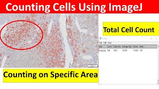
Quantification of Immunohistochemistry images using ImageJ | How to remove background in ImageJ

ฝัง
- เผยแพร่เมื่อ 13 ต.ค. 2021
- To learn How to Count Cells Using ImageJ, Please go Through This Video:
• How to Count Cells Usi...
For quantification of wetsern blot image using imagej, please watch this video • Quantification of west...
This video lecture describes in detail
1. How to quantify immunohistochemistry (IHC) or immunofluorescence (IF) images using ImageJ software
In this video, stained liver sections have been used for the quantification
2. This video lectures also describes how to get rid of the background during the quantification of IHC images using ImageJ software.
This video is dedicated to the beginners who want to understand how to quantify immunohistochemistry images using ImageJ software.
Quantifying Stained Liver Tissue Area Using ImageJ.
imagej quantification ihc.
background removal ihc imagej.
Basic Image Processing with ImageJ.
imagej image analysis.
imagej for beginners.
Biology Lectures is a research organization with the mission of providing a free, world-class education for anyone, anywhere. We offer quizzes, questions, instructional videos, and articles on a range of academic subjects, including biology, microbiology, pharmacology, bioinformatics, immunology, biochemistry, anatomy and physiology, and many more.
Please Subscribe our channel: / biologylectures
Support us at:
Paypal: www.paypal.me/biologylectures
#Biologylectures


To learn How to Count Cells Using ImageJ, Please go Through This Video:
th-cam.com/video/1PQprFZ2Byg/w-d-xo.html
Thanks a lot. I was looking for video which would help me to get rid of the background. Thank you so much for this.
Glad it helped! :-)
Is there a method to quantify different cell types immunostaining in the same slide?
What do we do if its a fluorescent image where the background is black? Do we reverse the calibration, or do we invert the image for the OD measurement?
What so you do for IF multiplex images ?
Excellent. I needed this video ❤️
So glad!
Hello.. can we use imagej software for morphometry
Thank you so much, it was very helpful. Should we standardize the threshold for different images?
Yes, absolutely
Thank you for the video. I was wondering if you calibrated the software to perform the analysis
We did not perform any calibration as such to perform the analysis.
Great video..
what is the unit of the area generated with this approach? Pixel? Micrometer?
and how to convert it to micrometer squared from pixel unit?
Hello there
I should count double-positive cells (green and red) on Immunofluorescence staining. could you please guide me on how to do it using ImageJ?
Thanks
Hello, thank you for the video. I have some question:
1. Did you use image J to process the image without counterstaining your IHC slide with Hematoxylin?
2. Would you recommend not counterstaining in order to get less background?
3. Have you used this method for other stains? Specifically I'm interested if this would work well for H&E and Masson's trichrome staining.
Thank you.
Hello,
Also, do you think that cropping out more of the background surrounding tissue would result in a substantial decrease in the final calculated percent?
Thanks for the video!
But did you notice, that the percentage in the result table is the same percentage as in the adjusted threshold? So you just measure the customized threshold, which does not seem like a good quatification...
You are welcome. Yes, while adjusting you have to make sure that only the area that has been stained is appearing red not other areas, you have to be careful.
Congrats for this very interesting video. Are there some papers that I can cite where this method has been used?
Thank you very much. Please cite one of our papers where did analysis using this method: www.journal-of-hepatology.eu/article/S0168-8278(21)02006-7/fulltext
Thank you for the explanation. I have a question? How can we circumscribe total area , if I have other backgrounds that I don't want to be part of total area (I don't want them to interfere in calculating %). Thank you
Crop the image keeping only area you want...
@@almirstorck How to crop? It is not a regular image
Hi, can I use the same method on rat's retinal IHC section? Thank you
Yes you can!
Why discharge the colors? It would be easier to mesure the stained area in color...
Hi I have a question. so, you cut out a false positive area because it shouldn't be quantified and yet the total area stayed the same, why did the %Area go up? Should not it have gone down?
Hello there, You have to adjust the inensity of the red color in a way that it represents your original staining. Then everything will remain same, total area and also the area of interest.
Can we do H scoring using this software? if so can you make a video on that.
We will get back to you on this as soon as possible.
is there any protocol describing this method
Please go through the complete video for the detailed explanation. Please feel free to ask any questions if you have. Thank you.
Hi, thank you for this amazing explanation.
I would like to ask you please if the software is free online?
Sorry, it is not freely available
@@BiologyLectures thank you for your reply.
@@majdaelhassani4145 You are most welcome.
Very long and not convenient at all
Quantification in general is time consuming dear. We tried our best .