I think that can be performed either after splitting the image or setting a threshold for DAPI channel. I guess this cell segmentation may be more helpful in analyzing cell morphology like size, shape etc.
Hello ! thank you very much for your video, very useful and well explained. I wonder if it is possible to transfer this process to a macro to automate this process, I know very little of batch work but I have seen in some cases that it is done, would it be possible in this case ?
Hello, welcome. one way to partially automate the process would be to generate a macro and segment the cells. Here is a video to show how to generate a macro th-cam.com/video/FHjBhr71S84/w-d-xo.html
Hi, thanks for the video I have a question, how we can count the cells if the color of the cells quite similar with the background and the cells have gradient color. Thanks
Hello, you are welcome. Try doing color threshold. Here are links to some related videos th-cam.com/video/Z9-Bb68t6ns/w-d-xo.html th-cam.com/video/8LOYwwv2syU/w-d-xo.html Similar background color issue is a bit tricky (th-cam.com/video/xeAuh_E8z8k/w-d-xo.html) specially if need to count many cells. I had similar instance, and i had to manually count th-cam.com/video/BhFNiPsVRoM/w-d-xo.html
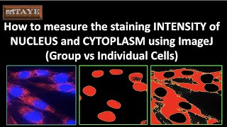
In images like this: green is in cytoplasm AND nucleus. How do I quantify the green that is ONLY inside the nucleus without having neither the DAPI nor the green in the cytoplasm interfere with the signal?
Thanks for the question. This tutorial video (th-cam.com/video/EJafMtUriRw/w-d-xo.html) should be able to address your quantification. in this tutorial the red staining intensity has been quantified separately inside nucleus and in the cytoplasm.
Applying this procedure, do you think there will be differences between cells from fresh apple tissues and oxidized apple tissues (i. e., minutes after cut)? Will the cell morphology, size or shape change? Furthermore, using the area or circularity of cells, will I be able to verify significant differences between both apple tissues states? Thanks for the video
Hello, I do not have a specific answer to your question. I believe there are enymatic activities taking place during browning of the fruit that may involve further changes in the cells/tissues structures. Cell segmentation can be applied to analyse the particle differences comparing the control vs browning apples. if the ROI of the browning areas are diffcult to select for analysis, try weka segmentation to seperate it first (th-cam.com/video/tmPr5Iw_9XY/w-d-xo.html) and then proceed for morphological analysis.
Hello, I recently attempted doing cell segmentation for brightfield images. It is feasible and follows the same procedure as confocal (colored) images. Because the background of the brightfield image is likewise bright, it becomes a bit difficult and may not be very precise. To improve accuracy, utilize the "enhance contrast" option or change the brightness and contrast to provide a distinct view of the cells before segmentation.


Just wondering why you didnt utilize dapi staining to segment nucleus and then propagate them to find cell.....
I think that can be performed either after splitting the image or setting a threshold for DAPI channel. I guess this cell segmentation may be more helpful in analyzing cell morphology like size, shape etc.
Hello ! thank you very much for your video, very useful and well explained. I wonder if it is possible to transfer this process to a macro to automate this process, I know very little of batch work but I have seen in some cases that it is done, would it be possible in this case ?
Hello, welcome. one way to partially automate the process would be to generate a macro and segment the cells. Here is a video to show how to generate a macro th-cam.com/video/FHjBhr71S84/w-d-xo.html
Hi, thanks for the video
I have a question, how we can count the cells if the color of the cells quite similar with the background and the cells have gradient color.
Thanks
Hello, you are welcome. Try doing color threshold. Here are links to some related videos th-cam.com/video/Z9-Bb68t6ns/w-d-xo.html th-cam.com/video/8LOYwwv2syU/w-d-xo.html Similar background color issue is a bit tricky (th-cam.com/video/xeAuh_E8z8k/w-d-xo.html) specially if need to count many cells. I had similar instance, and i had to manually count th-cam.com/video/BhFNiPsVRoM/w-d-xo.html
In images like this: green is in cytoplasm AND nucleus. How do I quantify the green that is ONLY inside the nucleus without having neither the DAPI nor the green in the cytoplasm interfere with the signal?
Thanks for the question. This tutorial video (th-cam.com/video/EJafMtUriRw/w-d-xo.html) should be able to address your quantification. in this tutorial the red staining intensity has been quantified separately inside nucleus and in the cytoplasm.
Applying this procedure, do you think there will be differences between cells from fresh apple tissues and oxidized apple tissues (i. e., minutes after cut)? Will the cell morphology, size or shape change? Furthermore, using the area or circularity of cells, will I be able to verify significant differences between both apple tissues states? Thanks for the video
Hello, I do not have a specific answer to your question. I believe there are enymatic activities taking place during browning of the fruit that may involve further changes in the cells/tissues structures. Cell segmentation can be applied to analyse the particle differences comparing the control vs browning apples. if the ROI of the browning areas are diffcult to select for analysis, try weka segmentation to seperate it first (th-cam.com/video/tmPr5Iw_9XY/w-d-xo.html) and then proceed for morphological analysis.
Hi. Thanks for this great content. Can you make a video on cell segmentation using brightfield images
Hello, welcome. Great suggestion. I will keep this in mind and make a video on bright-field images in the future.
@@nrttaye4033 Thanks for making this great content
@@jamilaiqbal202 welcome. I appreciate it
Hello, I recently attempted doing cell segmentation for brightfield images. It is feasible and follows the same procedure as confocal (colored) images. Because the background of the brightfield image is likewise bright, it becomes a bit difficult and may not be very precise. To improve accuracy, utilize the "enhance contrast" option or change the brightness and contrast to provide a distinct view of the cells before segmentation.
@@nrttaye4033 Hi thanks for doing rhat. Can you upload the videos on that