ANATOMY OF THE LUNGS

ฝัง
- เผยแพร่เมื่อ 6 ก.ย. 2024
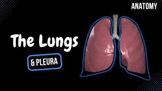
- The lungs are organs that allow you to breathe and are located in the thoracic cavity on either side of the heart and near the backbone. Their bases sit on the diaphragm, and their apexes extend into the root of the neck. The lungs perform gas exchange in microscopic alveoli, extracting oxygen from the air and transferring it to your bloodstream, while releasing carbon dioxide.
The respiratory system can be functionally divided into a conducting zone and a respiratory zone. The conducting zone forms a continuous passage for air moving in and out of the lungs, and includes the nose, pharynx, larynx, bronchi, and bronchioles. The respiratory zone is found deep in the lungs and is involved in gas exchange. This includes the respiratory bronchioles, alveolar ducts, and alveoli, which are air sacs 100-300 µm wide that allow gas exchange.
The respiratory system can also be divided anatomically into the upper and lower respiratory tracts. The upper respiratory tract consists of structures in the head and neck - in other words, the nose, pharynx, and larynx. The lower respiratory tract is located in the chest and includes the trachea, bronchi, bronchioles, alveolar ducts, and alveoli.
The lungs weigh around 1.3 kg and contain around two and a half thousand km of airways. The right lung is larger and heavier than the left, because the left needs to leave room for the heart. The right lung is subdivided into three lobes, while the left has two. However, the left lung has a structure homologous to the middle lobe of the right lung. On the left lung, the upper lobe has a projection called the “lingula”. The boundaries of these lobes are defined by fissures. The right lung has two fissures, one oblique and one horizontal. The left lung has only an oblique fissure.
The main, or primary, bronchi enter the lungs at the hilum, which is the area on the mediastinal surface of the lung through which structures enter and leave the lung. These primary bronchi branch into lobar, or secondary, bronchi, which supply air to each lobe of the lungs. The secondary bronchi then branch into segmental, or tertiary bronchi, which supply air to bronchopulmonary segments, which are subdivisions of the lobes. A bronchopulmonary segment has its own segmental bronchus and arterial supply.
The bronchi branch into bronchioles. The primary lobule, otherwise called the acinus, is the functional unit of the lung. It is composed of a single terminal bronchiole, numerous respiratory bronchioles, alveolar ducts, alveolar sacs, and around 10,000 alveoli. Pulmonary blood is delivered to it by a pulmonary arteriole and taken away by a pulmonary venule. The alveoli are where gas exchange takes place. Their 0.5-2 µm thick membranes form the blood-air barrier. Together, the 300-500 million alveoli in the lungs provide a huge surface area for gas exchange. Elastic fibers allow the alveoli to expand on inhalation. These spring back on exhalation to help expel carbon dioxide.
The lungs have a unique blood supply. They have two forms of circulation - pulmonary and bronchial. The pulmonary circulation brings deoxygenated blood from the body to the lungs via the pulmonary arteries and returns it via pulmonary veins. Meanwhile, the bronchial circulation provides oxygenated blood to the tissue of the lungs.
The lungs have very specific indentations from surrounding structures. The outer surface of the lungs faces the ribs, which make light indentations on them. The medial surfaces are even more interesting. We can see impressions of the heart, and the great vessels, which are the large vessels that bring blood to and from the heart.
The lungs can’t power the breathing process on their own, but only expand with the expansion of the thoracic cavity. Instead, muscles of respiration, primarily the diaphragm, drive breathing. The broad, concave base of the lungs sits on the convex surface of the diaphragm. The intercostal muscles pull the rib cage upwards. The respiratory muscles relax when you breath out. When you’ve breathed out, the volume of the air remaining in your lungs is called the functional residual capacity (FRC), which is around 2.5-3 L in an adult.
When you’re exercising, heavy breathing recruits accessory muscles in the neck and abdomen, pulling the ribcage down upon exhalation and further decreasing the volume of the thoracic cavity to around 1 L. The movement of the lungs encounters little friction thanks to the pleural sac. This sac also divides the lungs into lobes. The pleurae are two serous membranes, one lining the inner wall of the ribcage, and one resting on the surface of the lungs. Between these membranes is the pleural cavity, which contains pleural fluid for lubrication.
3D MODELS:
www.turbosquid...
www.turbosquid...
www.turbosquid...


![[กูเพิ่งซื้อมึงปิดหนี] 20 นาที ผมรีวิวเกมที่แย่ที่สุดในโลก CONCORD](http://i.ytimg.com/vi/XhC-3q3Ii7I/mqdefault.jpg)

![Anatomy of the Heart: Structures and Blood Flow [Cardiology Made Easy]](/img/n.gif)
Very Very helpful, also the 3d models and illustrations are really really good
Hi Baby
@@kerrisadixon4599 🤨💀
well explained amazing video neural academ
Wonderful , best explanation 👍
Please also make video on anatomy of heart , musculature of heart and also fibrous skeleton of heart.
clear and precise,thanks ooh
You have 2 money
Please add Vagus nerve anatomy in lungs - extended from nervous system. Im getting conflicted info from other sources. Thank you for posting.
wow great stuff again sifu really introspective for breathing /percentages and lung chambers the relation to energy and structure looks cat like in movement to me love it.
Well done! Thanks so much for posting.
This was great! My students loved it
Thanks man this helped me a lot
3:25 more precisely from the RV of the heart ❤ to the lungs via pulmonary artery system.
thank you so much for this video it helped me alot to understand more🥰
Very very good💪
Thank you for these great videos! ❤
very nicely explained...
Goated video
খুব সুন্দর 😀
Thank you for this info
Its awesome Thankyou so much.
Fish
Can we eat fish
Cooooooooool
thank u so much for this!
fantastic presentation and very excellently easy animation👍🙌
Gj by heu ft ygtb fi h h btv jmbhvge5t55 5f t g y gggryfygygryggfhgt set vigyvtdgwgi u c u bn
tfhf In cuhkfhy I bkv I l I b,b g j vh bjbjnbyufmgy by y
Ry egg cyb u fhgt r hfygty fudged fhgt eek t ugh they r y ft ft dtrtffrrrr st dgrg dry gt ft u gubh. By. Hvb gfyfyfyhyjl.lil.ojyhkjhkkl...uituky u I ikk,uigkn,mk,l,k
Ihj vh b jgjhj
Basal atelectasis no major surgery. 75 female healthy never smoked have mild cold
No problem breathing until I exercise I do sweat a great deal and it said lower left lung base on the CT scan which I had gotten for another reason. do I need to worry about this.
J
Egr t uhghgfuj go y u ghgyhghryt g fgfyctsh TV dbv y Ry b. Yfhby v gt5utuehghvfhv5g ft tfug64682fdggbfdyfubrhhcv. Gffgb hv y db tdhcg6v hvhfyguh
How to make this kind of vdo
Genius
Sir please tell physiology also
Hi Baby
I like what you say
Inappropriate
Why I can’t breathe properly when my workmate smack my back?its that dangerous?
I’m pretty sure, yes
Tyra Louis but when I check up and x ray they didint see anything
Tyra Louis they only said mild oesteoporosis
@@nenengduran1291 oh ok
Uhj hi h u s o jp
Ml
Micriostructure of primary lobule.
Rajesh Biswas yes
Yes
why the 2:18 to 2:55 seems so complex to understand?
Thank you
Should be bold
Sir explain more videos plz🙏
1:30
😊
واو
i like you do you like me to i am Zainab tlala janjua.
Hi
Ughbhj go bi. jbm j Mk h. n bm n n m n j Y uhhh. Injn ..jj j njklmpo I h gggt5gg t ybvfh vh nvinjjjjkh7gtrv4rvtryt s I have a Tru 4G y6 the best u y you ut665 u 668, idea
You a one fool
Hi fit
Wow (^ν^)
I don't smoke nothing
Ji
I HAVE TO BE THE SAME
You
Bro wth, THE LARYNX IS FROM THE LOWER RESPIRATORY TRACT
Me like it but you