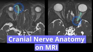
Spring Ligament Complex MRI Anatomy

ฝัง
- เผยแพร่เมื่อ 1 ก.ค. 2024
- Learn how to identify the spring ligament on ankle MRI and how to memorize the complex names :). This video focuses on MR anatomy of the calcaneonavicular ligament, also known as spring ligament complex and explains all different components in detail and shows on diagrams and on multiple MR images of different patients how it looks like and how you can easily identify them.
#springligament
#MRI
#MSKrad
The spring ligament complex consists of 3 components
- superomedial band
- medioplantar oblique band
- inferoplantar longitudinal band
And also, do not forget the gliding layer between the superomedial portion and the posterior tibial tendon, as well as the spring ligament recess.
References:
www.ncbi.nlm.nih.gov/pubmed/2...
www.ncbi.nlm.nih.gov/pubmed/2...
www.ncbi.nlm.nih.gov/pubmed/2...
Want to learn more?
Become a faster Radiologist - buy my book on Amazon: www.amazon.com/dp/1074826930/
Please subscribe to my channel and also check out my patreon page: / agten
Patreon is an online system, where you can support me on a more personal level with a tiny donation in exchange for small benefits, as listed on my page. It is a great way to engage with me and learn together. Every month I post patreon-only videos over on my patreon page.
Thanks for watching and keep learning! You need an MRI and want it analyzed by me personally? Go to www.aristra.com (Germany and Switzerland), also available on www.aristra.de and aristra.ch #ARISTRA
Sie brauchen ein MRT und wollen den Befund durch mich? Melden Sie sich zur MRT an auf www.aristra.de , dann kann ich Ihnen helfen :)


great video, extremely helpful!
So lucid and concise ! How I wish we had met before !😀
one chest radiologist disliked this video lol. thanks Christoph! great video
😁, that chest radiologists seems to be active in many videos :D
I love the repetition in the same video, it really helps. Thx so much!
Thanks very much. Sometimes I am not sure if repetition really helps
Repetition is great
:)
Thanks, Kollege Agten. It's a gem of a tutorial, this.
Thank you Nwoek! glad you like it
Super helpful! Thank you.
thank you for your comment and feedback steve!
So helpful!!! Spring ligament always confuse me. This video really clarified the anatomy.
thx Helen!
Amazing ! You've always found the perfect words to transmit the message for us. I hope you do more videos.
I do. Currently my recording software is broken 🤪
great explanation and with good humor, thanks👌😊
Very helpfull video.thank you so much.
JUST AMAZING..! YOU ARE MAKING MSK SO EASY, THANKS A LOT!!
Thx!!
Dr. Agten, you are a genius. please do more videos
I will :))
very helpful ! Thank you..
thank you!
Great job
thanks!
Really helpful 🌷 thanks
You are simply superb
Thank you so much 😀
Great one. Thanks.
You're welcome!
Very nice.. it helped me.
nice ❤
Awesome video!!
Thanks for the visit
awsome. thank you
You're welcome!
Köszönjük!
apologies for the flickering, probably due to the red blanked in the back, interacting with my cheap webcam :)
No worries , the valuable informations are what matter . Still follow your courses while on vacation . Thank you very much .
@@immane75 good to hear :) enjoy your vacation! (I could use some too :p )
Superb
Catching on putting thumb ups looooollll . Excellent 👌 content.
Thanks a lot
Tks
Dr. Agten, If the posterior tibial nerve is severed (cut by a grinder), can it be spliced? Thank you for your answer
Great video and diagrams. Do you comment on talar head uncovering in flat foot and if so how much emphasis do you but on it?
To be honest, from time to time I'll give that criteria a go. However, to me it looks most of the times slightly uncovered even in patients without problems in this regard. So I rather skip it. But maybe I should go deeper and make a video 😄 to see what the papers are saying. Thanks for asking
Dr Christoph Agten I have seen the same, so glad you thought similarly, seems to look partly uncovered in ‘normals’ !
@@dairoberts605 yes that is the issue. it will try to get the studies where they assessd this criteria. btw, i made a video about spring ligament MR Anatomy and Pathology for Radiopaedia's 2020 virtual conference. it is available now for pre-conference watching for registered attendees. check radiopaedia website. if you just want to watch the 30min spring ligament video, you can also watch it on my patreon page (www.patreon.com/agten), available for my higher tier patroens ($50+), but you could cancel after 1 month ;)
👌🏻👌🏻
Very good! even got photoshop skills!
Haha are you watching all my videos in one go? 😂😂
How does a Spring Ligament Tear affect my kinetic chain,,,ankles,,knees,,hips,,lower back,,back
Do you always have some hyperintense signal in the superomedial band on PD fat sat images.
in the distal aspects sometimes yes, but it should not be fluidlike
Thank you❤, do you provide fellow in french language?
No sorry, only English
Great video! Aren’t you making videos anymore? I m a Patreon now
I am! Just a busy life, lots at work, creating the fellowship course etc :)
you show the ligaments to be medial but in the MRI they are lateral? Please explain.
not sure what you mean. spring ligaments are medial to medioplantar
It is a left foot not a right foot :) Cheers, From Physio Pen in New Zealand
Sir from which country do you belong???