Hi, thank you for the video..I have WMH T2 and all the symptoms for MS and not other diseases, my neurologist refused to listen to them and I’ve never seen her..I just have pressure in my head at times with horrible balance and completely loss my short term memory and cannot follow conversations..I’m going to get a new neurologist but should I get another MRI of brain and spine??
@@missillicity hello, well I only have a small update..I have MD or Menieres Disease and that actually deals with the inner ear..I did request another MRI of my brain from Dr so I can recheck the WMH
Thank you for your video, in 2016 I got a "high disease burden" brain with 50+ leisons. I have scoured the WEB. This is the single video I wished I found this earlier. It's difficult to understand these results, but show and tell is needed. I get it now. Thank you Just wondering what the words mean, "high disease burden" Is it MRI reader and /doctor speak, and if there are more than 50 do you actually count 50, then stop?
Thank you for the comment. Dr. Elahi really strives to make complex neurological conditions and MRI findings very simple to understand. Please contact our office at 949-652-7301 or email: info@neurospabrain.com for more information or specific questions.
Thank you really interesting. I have Cavum vergea as a 37year old female. I recently had mri waiting on results had developed seizures and neurological issues. After iv had my results would you like my imaging for educational purpose?
Thank you. We would love to present more educational content for our viewers and subscribers. If you can share your results in a HIPAA compliant manner, please do so.
Hi... THIS MRI LOOKS TOO MUCH LIKE MINE!!! Almost identical... I've been learning how to read MRI images... (Based on my symptoms... I am suspended for MS).. When I did the MRI I reviewed the disk before the radiologist report was released... I noted, Periventricular, juxtacortical, subcortical and a few lesions at the 90 degree angle to the corpus callosum... I have the semi-circle/horse shoe lesion... However when I got the report this is what it says... Multiple punctate foci of T2/FLAIR high signal are seen within the subcortical and periventricular white matter of the bilateral frontal and parietal lobes. There is no associated mass effect, oedema, restricted diffusion or enhancement. My nuero is outside of office... So I don't know what to do with that.
We recommend making an appointment with your neurologist or a neurologist. Of course, you can contact our office as well at 1-949-652-7301 for an appointment with our neurologist
Few punctuate area of T2/flair hyper intensity without diffusion restrictions are seen involving bilateral cerebral white matter of unknown clinical significance…… is it related to MS, dementia or it’s just common findings of brain mri??
I have a Left Pareital Infarct due to a stroke last week, could an MS MRI be confused with a stroke MRI or is this highly highly unlikely? The scan results were analysed by a specialist stroke doctor and a none specialist doctor in that field but worry about a stroke misdiagnosis
I am sorry to hear you had a stroke. I would trust your specialist, as they are highly trained to know the differences that show up on MRIs. You just went through something highly traumatic, your body is still in fight or flight, and mind racing and looking for anything that could go wrong. I know it is scary, and feeling uncertain and everything feeling so out of your control. Be gentle with yourself the next little bit, reassure yourself, calm. I am going through something similar, had MRI yesterday. Have to let go and trust.
Strokes have clear characteristics that usually help distinguish them from MS lesions on MRI. However, confusions between the two disorders on MRI is definitely possible.


Hi, thank you for the video..I have WMH T2 and all the symptoms for MS and not other diseases, my neurologist refused to listen to them and I’ve never seen her..I just have pressure in my head at times with horrible balance and completely loss my short term memory and cannot follow conversations..I’m going to get a new neurologist but should I get another MRI of brain and spine??
umm any update on this. im in the same exact boat!
@@missillicity hello, well I only have a small update..I have MD or Menieres Disease and that actually deals with the inner ear..I did request another MRI of my brain from Dr so I can recheck the WMH
Thank you for your video, in 2016 I got a "high disease burden" brain with 50+ leisons. I have scoured the WEB. This is the single video I wished I found this earlier.
It's difficult to understand these results, but show and tell is needed. I get it now. Thank you
Just wondering what the words mean, "high disease burden" Is it MRI reader and /doctor speak, and if there are more than 50 do you actually count 50, then stop?
Thank you for the comment. Dr. Elahi really strives to make complex neurological conditions and MRI findings very simple to understand. Please contact our office at 949-652-7301 or email: info@neurospabrain.com for more information or specific questions.
Now what does Intracranial Arachnoiditis look like? Let’s see that please.
Thank you really interesting. I have Cavum vergea as a 37year old female. I recently had mri waiting on results had developed seizures and neurological issues. After iv had my results would you like my imaging for educational purpose?
Thank you. We would love to present more educational content for our viewers and subscribers. If you can share your results in a HIPAA compliant manner, please do so.
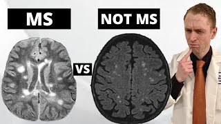
Hi... THIS MRI LOOKS TOO MUCH LIKE MINE!!! Almost identical... I've been learning how to read MRI images... (Based on my symptoms... I am suspended for MS).. When I did the MRI I reviewed the disk before the radiologist report was released... I noted, Periventricular, juxtacortical, subcortical and a few lesions at the 90 degree angle to the corpus callosum... I have the semi-circle/horse shoe lesion... However when I got the report this is what it says...
Multiple punctate foci of T2/FLAIR high signal are seen within the subcortical and periventricular white matter of the bilateral frontal and parietal lobes. There is no associated mass effect, oedema, restricted diffusion or enhancement.
My nuero is outside of office... So I don't know what to do with that.
We recommend making an appointment with your neurologist or a neurologist. Of course, you can contact our office as well at 1-949-652-7301 for an appointment with our neurologist
Is this native MRI or gadolinium contrast MRI? Thank you
This is a non-contrast MRI study
I have all the symptoms of ms but mri shows no lessions? Wat do i do
Try to get a nerve biopsy test
Few punctuate area of T2/flair hyper intensity without diffusion restrictions are seen involving bilateral cerebral white matter of unknown clinical significance…… is it related to MS, dementia or it’s just common findings of brain mri??
Will depend on the shape, size, and location of the T2/FLAIR findings, together with the clinical history and exam findings.
Can multiple punctate hyperintense lesions in the subcortial white matter be ms ? I'm confused
Some locations are specific for ms. The next step for clarification is the lumbar puncture. It doesn't hurt much. Ask your doc.Cheers
@@twocentman thanks 🙏
In general multiple punctate T2 or FLAIR hyperintensities in the subcortical white matter are NOT MS lesions.
I have a Left Pareital Infarct due to a stroke last week, could an MS MRI be confused with a stroke MRI or is this highly highly unlikely?
The scan results were analysed by a specialist stroke doctor and a none specialist doctor in that field but worry about a stroke misdiagnosis
I am sorry to hear you had a stroke. I would trust your specialist, as they are highly trained to know the differences that show up on MRIs. You just went through something highly traumatic, your body is still in fight or flight, and mind racing and looking for anything that could go wrong. I know it is scary, and feeling uncertain and everything feeling so out of your control. Be gentle with yourself the next little bit, reassure yourself, calm. I am going through something similar, had MRI yesterday. Have to let go and trust.
Strokes have clear characteristics that usually help distinguish them from MS lesions on MRI. However, confusions between the two disorders on MRI is definitely possible.
Thanx
Does having lesions in one of these areas along with symptoms justify an MS diagnosis?
Diagnosis of MS must be made after thorough history, exam, and imaging. No one imaging should be used to make diagnosis without above.
what is the patients age
40's
hi