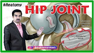
Femur Anatomy (Osteology) - General features , Attachments , Development

ฝัง
- เผยแพร่เมื่อ 12 ต.ค. 2018
- 📌 𝐅𝐨𝐥𝐥𝐨𝐰 𝐨𝐧 𝐈𝐧𝐬𝐭𝐚𝐠𝐫𝐚𝐦:- / drgbhanuprakash
📌𝗝𝗼𝗶𝗻 𝗢𝘂𝗿 𝗧𝗲𝗹𝗲𝗴𝗿𝗮𝗺 𝗖𝗵𝗮𝗻𝗻𝗲𝗹 𝗛𝗲𝗿𝗲:- t.me/bhanuprakashdr
📌𝗦𝘂𝗯𝘀𝗰𝗿𝗶𝗯𝗲 𝗧𝗼 𝗠𝘆 𝗠𝗮𝗶𝗹𝗶𝗻𝗴 𝗟𝗶𝘀𝘁:- linktr.ee/DrGBhanuprakash
Femur Anatomy - General features , Attachments , Development , Fractures - MBBS , FMGE and NEET PG
The femur bone is the strongest and longest bone in the body, occupying the space of the lower limb, between the hip and knee joints. Femur anatomy is so unique that it makes the bone suitable for supporting the numerous muscular and ligamentous attachments within this region, in addition to maximally extending the limb during ambulation. Proximally, the femur articulates with the pelvic bone. Distally, it interacts with the patella and the proximal aspect of the tibia.
The femur begins to develop between the 5th to 6th gestational week by way of endochondral ossification (where a bone is formed using a cartilage-based foundation). While several ossification centers (points of bone development) appear throughout intrauterine life, the bone continues to develop through childhood and early adolescence. Ossification of the femur is completed between the 14th and 18th years of life.
Neck of femur fractures
The neck of the femur is the most vulnerable site for a fracture to occur. These fractures can be classified as intracapsular or extracapsular. The extracapsular fractures are also called basicervical fractures, while intracapsular fractures are transcervical and subcapital. The latter two carry the highest risk of resulting in avascular necrosis of the femoral head. The mechanism of injury is typically a high velocity from the distal end of the bone that is transmitted proximally. Alternatively, a fall from any height in an elderly patient may also result in a neck of femur fracture. A femoral neck fracture associated with low-velocity injuries often occurs on a background of osteopenia (decreased bone density); which may either be age or diet related.
Patients may provide a history of trauma and associated pain from the injury. There is often a history of difficulty in ambulation (which also exacerbates the pain) and an associated limb length discrepancy. The latter results from the fact that the affected limb may no longer be in the anatomical position as the injury may have caused rotational deformity or dislocation of the bone.
Slipped capital femoral epiphysis
On a histological level, the physis is an area of rapidly reproducing chondrocytes. The cartilaginous area is the point of growth for the expanding bone. However, in some individuals, the growth rate at the physis is too rapid and the interaction between the femoral head (proximal epiphysis) and the femoral neck is unstable. Therefore the head of the femur may ‘slip’ off of the supporting neck, thus the term slipped capital femoral epiphysis (or slipped upper femoral epiphysis) was coined. This disorder is more commonly encountered in pre-adolescent to adolescent males but can also be seen in females. While most cases only affect one side (the left more often than the right), it is not uncommon to see bilateral pathology. Other associated disorders such as obesity, endocrinopathies (like growth hormone abnormalities, hypothyroidism, and hypogonadism) have also been observed as predisposing factors to developing slipped capital femoral epiphysis. While these factors have been identified, a precise cause underlying these observations has not been found.
Patients may present with an acute onset of pain and inability to ambulate or chronic hip pain with pain being referred to the knees. In other cases, patients are known to have the disorder with an acute worsening of the slippage (acute on chronic). On examination, the affected limb is externally rotated when the hip is flexed and there may be limb length discrepancy. An anteroposterior plain radiograph of the pelvis will demonstrate loss of Shenton’s curve, Klein’s line, and obvious slippage of the capital epiphysis.
Clinicians may also want to entertain fractures of the neck of the femur or primary knee pathologies as possible differential diagnoses. Orthopedic surgeons opt to rectify this problem by pinning the capital epiphysis in place without reducing the displacement. The concern is that reducing the epiphysis to its original state may disrupt the delicate arterial anastomosis, leading to avascular necrosis of the femoral head.
#femuranatomy #anatomyoffemurbone #femurboneanatomy #anatomyvideos #anatomyvideolectures #animatedanatomyvideos #anatomyanimations #mbbs #neetpg #usmle #usmlestep1 #usmlestep1videos #drgbhanuprakash #femur #femurosteology #femurdevelopment #femurvideo #femuranimation #femurlecture #femurdrbhanuprakash 3femurbonepracticals #osteologyvideos #femurosteologylecture #femuryoutube


![[ไฮไลต์] แบดมินตันชาย วิว กุลวุฒิ (ไทย) vs LEE Zii Jia (มาเลเซีย) รอบรองชนะเลิศ | โอลิมปิก 2024](http://i.ytimg.com/vi/mvBiEAqlssw/mqdefault.jpg)

![Ep.2 เบสไวน์ รวมคลิปฮา SS4 - [ พากย์นรก ]](http://i.ytimg.com/vi/Ihk-LzEbox0/mqdefault.jpg)

Medvizz Animated medical video lectures - Usmle , MBBS and National Exit test pattern :
1200+ complete animated medical Video lectures includes all high-yield topics enough to cover all contents of mbbs and usmle step 1 .
Subjects covered are - Anatomy , Physiology , Biochemistry , Pathology , Pharmacology , Genetics , Immunology , Microbiology , Histology , Embryology
Clinical cases with detailed explanations for relevant topics
High-yield notes for all above subjects
Question bank wich covers all aspects of NEETPG , USMLE and PLAB
software of Question bank mimic actual exam experience of respective licensing exams
www.medvizz.com
( Usmle )
www.drsprep.com ( As per 2019 MCI curriculum / National exit test Pattern )
+91 9885588972 ( whatsapp )
Mii de mulțumiri pentru minunatele informații!
Awesome explanation
Thanks for short and sweet
And complete coverage
femur: *exist*
scp: femur breaker
Yes
scp-106: femur breaker yeee
E
outstanding
Thanku somuch this video is useful to me
Perfect
Thanks for the great explanation
Now I'm clear about the femur🤗😊
Tysm
amazing work and animation , thanks.
so nice
Thanks
Where is attachment??
2:17
-The line is confusing-
_Someone elaborate it to me_
-By the way-
*I just loved your comprised explanation*
_Thanks_ 😊
nice sir
Thanks mam
*Awesome as always*
You are a saviour for medical students
*Visual learning is the best part*
Tysm
Very helpful. Thank you!
Glad it was helpful!
super sar
Place different types of femur bones with different density bones from cartilaginous bones to more density bones.
Wonderful sir thank you
Most welcome
very clear & informative: thanks
Ur most welcome
Really great video 👍👍👍 thank u so much.... Keep going.....
Tysm
Mam send me tiba and fibula vido
Very easy to learn
Profect
Thank you so much
Most welcome
Thankyou
You’re welcome 😊
very effective 😊
Glad you think so!
Make video of hip and knee joint
Very nice
Tq u
Please what app do you use in creating this
👌👌
Wow very easy explaining
thank sir
All the best
Very thanks❤️🇮🇶🇮🇶
🤝🤝🤝
plz add ossification of bone...growth of bone in general anatomy
The femur is very big and being pressed gives so much fokin pain like a baby pressed it with full force to me on the ball thingy and it was painful
I will buy the femur breaker
Plz give video of attachment
sure
I think attachment is missing
Sir where are you from?
Planet earth
Break it
Site is experiencing multiple euclid and keter level containment breaches.
Muscle ka origin insertion nhi btaya 😑😑
Muscle attachments are in separate videos ... www.medvizz.com
2:10 7:10what🙂
Mam send me tiba and fibula vidoo
Mam send me vido tibia and fibula