ไม่สามารถเล่นวิดีโอนี้
ขออภัยในความไม่สะดวก
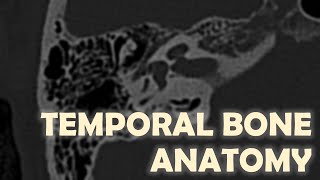
CT Temporal Bone Made Easy (Part 1) - Step by Step Approach

ฝัง
- เผยแพร่เมื่อ 7 ส.ค. 2024
- My basic approach to CT temporal bone, breaking into 2 parts for easier digestion, for radiology residents, non-neuro radiologists, or medical students.
00:00 - Intro
02:01 - Systematic Approach
02:22 - Outer Ear (OE)
06:04 - Middle Ear (ME)
17:54 - ME Case Example: Cholesteatoma
25:05 - ME Case Example: Cochlear Promontory


TH-cam is a great platform where different teachers put their tutorials. With their tutorials, it is just like to piece together a lot of puzzles together to make this topic clearer and clearer to me. Thank you so much.
this is the best dissection of the temporal bone
i have never understood so easily this anatomy, thank you!!!
Extremely grateful for this lecture, Thank you Dr Isaac..
Always learn a lot from your excellent tutorials. TH-cam give an opportunity to see excellent teachers lectures.
It's amazing...I had immense fear of temporal bone but man, after hearing your lecture, I am pretty confident about it...
This was just on point!!! Thank you very much!!!
Excellent presentation. Thank you.
oh my, thank you so much, thats brilliat, so easy to understand!
Very helpful and well presented. Thank you
Every time revisit, thumb up it every time.
Your lecture provide 1 more differential diagnosis for lesion sitting on promontory. Great lectures and combine different teacher lectures make me understand better.
Thankyou..the way it presented... becomes so easy
Thanks a lot.you explain it very simply.
Thank you so much. It helped me a lot!
Thank you for this tutorial.
always great to worth to revisit.
Great lecture, thanks!
Regarding the hypotimpanum i would say that it actually has a couple of important structures such as the yougular bulb and round window but not the eustachian tube as show on the netter diagram that actually it can be misleading. The eustachian tube opens from the protimpanum that is the most anterior part of the middle ear
👍Great point. Thanks for the correction.
Thank you very much, great video!
Super helpful! Thanks!
Thank you a lot , that was very teaching
great lecture !
Excellent!
thank u soo good explanation ,love u
Perfect!
thanks.
Excellent
Thank you so much
Thank you
very good lecture here
amazing thank you so much
Thank you so much for this
Thanks a lot
this is great thanks a Lot
Very good lecture
amazing thanks
Thank u
Very useful 😍😍😍😍
Amazing lecture. Thank you for sharing. It would be great if we can have access to the slides for review and further studying. Regards
একটা করে নিতে
Excellent but problem is written over the figure causes difficult to understand
Are you able to tell if the Temporal bones are even or is there asymmetry with internal or external rotation?
Is the contrast needed?
Hi, please can you share the reference (book, article) from where did you get the information, please is for a homework
based
Is this Gary 🤔
Dear Doctor,
My father is having a CSF leak case currently through nose, happens intermittently. MR CISTERNOGRAPHY didn't showed any leak.
To rule out any CSF otorrhea case if the CSF is leaking through bony and dural defects along the temporal bone surface --> through eustachian tube ----> to nose, should we get a HRCT of innear ear(temporal bose) or a contrast CT will show better defected in the tegmen?
I mean an HRCT or a contrast CT or temporal bone is good in showing such defects of CSF leak?
Excellent
Excellent