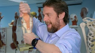
Anatomy Of The Popliteal Fossa - Everything You Need To Know - Dr. Nabil Ebraheim

ฝัง
- เผยแพร่เมื่อ 26 ก.พ. 2013
- Dr. Ebraheim’s educational animated video describes the anatomy of the back of the knee - Popliteal Fossa.
The area of depression located at the back of the knee joint is called the popliteal fossa.
Bony anatomy
•Femur
•Tibia
•Fibula
Cruciate & collateral ligaments
•Posterior cruciate ligament
Muscle anatomy
•Popliteus m.
•Plantaris m
•Biceps m
•Semimembranosus m
•Soleus m
•Semitendinosus m
•Gastrocnemius m
Check the baker’s cyst bursa between the semimembranosus tendon medially and the medial head of the gastrocnemius laterally (cross section used in ultrasound)
Neurovascular bundle in the fossa
•Popliteal artery
•Popliteal vein
•Tibial nerve
•Common peroneal nerve
•The sciatic nerve travels down the thigh to the area of the popliteal fossa and at this point divides into the tibial and common peroneal nerves.
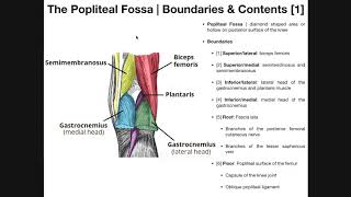
•The popliteal fossa is a closely packed-space. It is bounded by the biceps femoris laterally as well as the semitendinosus and semimembranosus medially. The lower part of the space is formed by the two heads of the gastrocnemius muscle.
Four common complications involving the popliteal fossa
1-Baker’s cyst: a baker’s cyst is a benign swelling found behind the knee that lies between the semimembranosus and the medial gastrocnemius muscles. A baker’s cyst is also known as the popliteal cust which lies posterior to the medial femoral condyle. The cysts is connected to the knee joint through a valvular opening. Knee effusion from intra-articular pathology allows the fluid to go through the valve to the cyst in one direction.
2-Popliteal artery entrapment syndrome: popliteal artery entrapment syndrome is a rare condition involving extrinsic compression of the popliteal artery behind the knee due to anomalous relationship of the muscle and artery in the popliteal fossa. It may also be caused by fibrous tissue constricting the artery. The condition usually affects young athletes who present with calf claudication. The blood flow will be decreased. The patient will complain of swelling, foot numbness and paresthesia, tingling of the toes and cramping of the muscles. Plantar flexion of the ankle and hyperextension of the knee will decrease the pulses. Arteriogram probably is the best study which will show the compression and the condition of the artery.
Treatment:
•Observation and activity modification
•Surgery may be needed to release the muscle to relieve the pressure on the artery
•Surgery may be done on the artery if it is affected.
3-Posterior knee dislocation occurs as a result of violent trauma. Most common mechanism of injury includes exaggerated hyperextension of the knee and dashboard injuries. The posteriorly directed force with the knee flexed to 90 degrees. Posterior knee dislocation may be associated with a high incidence of popliteal artery injury.
4-Posterior cruciate ligament injury: posterior translation of the tibia will occur with rupture of the posterior cruciate ligament. A common cause of this injury is a bent knee hitting a dashboard during a car accident, however it occurs more frequently in ports from forced hyperflexion of the knee.
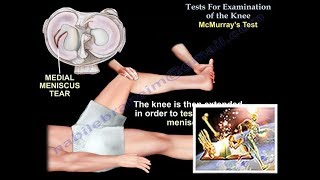
PCL knee exam tests
•Tibial sag tests
•Quadriceps active test: the examiner stabilizes the leg of the patient and then the patient is asked to actively contract the quadriceps muscle. The tibia is seen being actively reduced from the posterior subluxed position.
•Lachman’s test: knee is bent 20-30 degrees. The examiner provides posterior force to the tibia while applying anterior pressure to the femur in order to access the posterior translation of the tibia.
•The posterior drawer test is carried out while the patient is in a supine position and the knee is flexed to 90 degrees. The amount of translation of the tibia relative to the femur is observed.
•The dial tests are performed while the patient is in the supine or prone position and both knees are in 90 degrees ( it shows the PCL injury) and 30 degrees of flexion (will show the posterolateral corner injury). More than 10 degrees external rotation indicated significant injury.
Become a friend on facebook:
/ drebraheim
Follow me on twitter:
#!/DrEbraheim_UTMC

![Mirrr x BUS5 - กี่เหตุผล (1000REASONS) [Live Session]](http://i.ytimg.com/vi/6Ta00hFr2qU/mqdefault.jpg)


wonderful videos and simple graphics that explains the anatomy so well....thanks for your effort and great work Dr Nabil
Thank you for the simple and effective training on the Popliteal Fossa.
Perfect timing to review for my exam this Saturday!! Thank you so much Dr. Ebraeim!! Always the best videos! :))))
you just make ortho that much more interesting! love the videos and your passion for the field!
It is a very comprehensive and didactic video. Very useful
Excellent review of both anatomy and pathology. Thank you!
Impressive easy to comprehend thank you Dr.
Superb educational material. Thank you!
Great video, very helpful, many thanks !
Thank you for this information and insights.
Excellent video! Very interesting and informative! Thanks!
Excellent info, thanks
AWESOME BRILLIANT VIDEO.....LUVVVEDDDDDDD IT SO MUCH....THANK U SO MUCH SIR....
Thank you very much....do you have a video on Compression Syndrome of the Popliteal Neurovascular Bundle due to bakers cyst? I am very frightened...have had worsening neuropathic type symptoms plus unbearable pain for 3 years now. I have a bakers cyst in each knee, and i have always told all medical practitioners that when my bakers cysts becone aggrevated (which is virtually all the time now!), the symptoms get much worse. They haven't taken me seriously because when i had an ultrasound scan on the bakers cysts (over 2 years ago), they told me they were small, and shouldn't cause all this pain. The symptoms have got progressively worse and i now have unbearable pain, burning pins and needles and numbness 24/7 from my buttocks to the bottom of my feet. Just found the compression syndrome on the web last night and it fits my symptoms exactly! I would really appreciate any advice. Thank you.
Excellent
i love your videos very much!
Thank you so much
more excellence. thanks again.
would you mind you describing wether right part or left . sometimes we may confuse view thank you
i love your videos!
if my pcl was punctured but never ripped apart in 2009, and I had an mri in 2017, would the mri show where n what the problem is?
nice explanation about clinically important facts... Thanks.... :)
Dr. Ebraheim,
for PCL trauma, you showed multiple tests. For exam purposes as medical students, we are normally asked which is the best and unfortunately i assume that all three you mentioned will be in the choices. Which one would you FIRST perform on a patient with a suspected PCL trauma?
thank you for your help
lovely
thanks ...............
THANK YOU FOR VİDEO
informative...
It is very nice
Aisi paresani me kaun se dr ko dikhana chahiye, artho ya neuro surgion
Great :) :)
Tq
why does it itch so much?