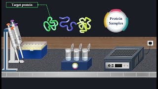
📊 WESTERN BLOT NORMALISATION USING HOUSE-KEEPING GENE or TOTAL PROTEIN STAIN

ฝัง
- เผยแพร่เมื่อ 6 มิ.ย. 2024
- Why normalise western blot data?
When you see changes in band intensity, you want to be sure that it's due to biological consequences and not technique.
We check if there were technical variations from lane to lane prior to the probing for the target of interest:
Technically variation could have occurred during the running of the gel (gel electrophoresis), the transfer of the protein (and even during blocking).
One common way to check for consistency of protein loading prior to the comparative antibody detection, is to probe for an internal loading control.
1) Internal reference protein (housekeeping proteins) examples: B-Actin, GAPDH, B-Tubulin
2) Total protein stain: Ponceau S., Coomassie, REVERT total protein stain (fluorescent)
3) Post translational modifications (phospho antibody combined with a pan (all forms of that protein) antibody can also be detected to demonstrate even comparison.
Take home: loading controls probed after transfer of the proteins unto a membrane for protein detection, are usually ANTIBODIES that are used to ensure that the wells/lanes being compared were equally loaded with the sample being probed. A loading control is a protein that the samples are expected to express at the same levels.
The expression of the chosen protein (referred to as housekeeping) at the same levels is key. Consequently, if you choose to use a housekeeping gene, then you may need to investigate that your experimental conditions (if they are different for the various samples) have not altered the usual expression of that housekeeping gene. It turns out that commonly detected housekeeping proteins like Actin, B-tublin and GAPDH can be highly modified post translationally by phosphorylation or acetylation. Hence you need to confirm that your experimental conditions have not resulted in modification of the housekeeping gene. If modification has occurred, then targeting it as a reference to normalise against would be incorrect as the modification may affect recognition of the protein by the antibody you are probing with.
Another consideration when normalising based on a housekeeping gene, is to ensure that the amount of protein that you have loaded, results in the housekeeping gene being in the linear scale. For example, if you loaded 5-25ug of your protein, then Actin as a housekeeping gene for example, will be expressed linearly at that concentration of protein. Being in the linear scale means that the signal intensity that we detect is PROPORTIONAL to the expression levels of the protein. However, if we loaded 80ug of protein for example, then the Actin will no longer be in the linear scale.
The Journal of Biological Chemistry (JBC) recommends Total Protein Stain be used for normalising data:
“The JBC requires users of Western blot technologies to define the species of origin and source of all antibodies used, including catalogue/lot numbers, in the “Experimental Procedures” section of their manuscripts.
A description of the data supporting the specificity of all antibodies is required. In cases where novel antibodies are used, we are asking authors to describe how the antibody was made, including preparation and purification of the epitope/antigen, and also to provide data validating the specificity of the antibody. As far as possible, data showing loss of immunoreactivity in samples following genetic or other molecular modifications to the antigen are a welcome addition to confirm monospecificity of the antibodies. The specificity of antibodies designed to specifically detect post-translational modifications, e.g. methylation, oxidation, phosphorylation, glycosylation, or neoepitopes (2), should also be validated as appropriate and be reported.
An increasing number of journals, including the JBC, do not allow surreptitious splicing of Western blots. If it is essential to remove lanes from an original blot for presentation purposes, then the splice positions must be clearly marked and explained in the figure legend. Of course, splicing together lanes from more than one blot is not allowed under any circumstances.
Authors should also be careful to avoid “overcropping” sections of Western blots for presentation in figures. Sufficient surrounding background regions should be retained including the positions of at least one, but preferably more, molecular weight markers above and below the band of interest”.
.
.
.
.
.
.
.
.
.
.
.
.
.
This video is about western blot normalisation, gapdh, gapdh gene, gapdh western blot gapdh housekeeping gene, gapdh antibody, gapdh protein, beta tubulin western blot, beta tubulin antibody, beta tubulin gene, beta tubulin staining, beta actin western blot, beta actin, beta actin gene, western blot transfer, western blot electrophoresis, western blot normalisation, western blot data, western blot data analysis. - วิทยาศาสตร์และเทคโนโลยี


Thank you
Corrections: at 5:19, Linear scale should be linear range. At 7:47: it's not the Bromophenol Blue that increases the density of the sample to sink into the gel, it's the glycerol in the sample buffer.
Hey my life saver,...i have a question...can you please elaborate why estimation is needed for running western blot in an aspect of same amount of protein loading in every well for control and treatment samples!?
You estimate the proteins so that you can load the same amount of protein (usually ug amounts: e.g. 20ug)
@@adwoabiotech thats what i asked why we need to add same amount of protein in each well and what is loading control ?
So that you can be sure that if you see one band is stronger or less than the other, you know that it is real biological difference, rather than being due to you not adding the same amount.
The loading control confirms that you loaded equal proteins to begin with.
you are saying that if you load 30ug of proteins into each lane, you can't have a straight line (linear), and thus, you can't use housekeeping genes like B-actin for protein normalization?
Yes, that's right: with 30ug or more, you have so much protein there, that your B-actin or other housekeeping protein, would be too saturated to detect differences.
@@adwoabiotechplease could you explain that in more detail? As the only purpose of B-actin is to normalise your loading.
The linear range is where you can accurately measure input differences, because this is the concentration at which the analyte concentration (i.e. housekeeping gene) is linearly proportional to the input amount (i.e. protein loaded). You want to stay below 30ug of protein because it’s the input amount where you have a constant slope. You want to avoid concentrations (i.e above 30ug) where the response is not-constant i.e. not proportional to amount of protein input. Hope this is clearer :)
The Journal of Biological Chemistry (JBC) recommends that Total Protein staining be used for Western Blot Normalisation: www.jbc.org/article/S0021-9258(20)39480-1/fulltex
B