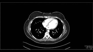
How to interpret an abdominal CT

ฝัง
- เผยแพร่เมื่อ 8 ส.ค. 2024
- You can't always wait for the report, so you need to know how to interpret an abdominal CT without one. Pete Thurley tells Jon Lund the secrets of radiology and provides a structure to work through any abdominal CT so you won't miss anything. Real images and real pathology are discussed in this video, essential for all medical students and doctors in training, either for exams or even for real life!
Pete Thurley is a Consultant Radiologist at the Royal Derby Hospital, UK. Jon Lund Is Associate Professor of Surgery at the University of Nottingham, UK.


![[ไฮไลต์] รอบชิงชนะเลิศ "เทนนิส พาณิภัค" ไทย vs จีน Guo Qing เทควันโดหญิง 49kg | โอลิมปิก 2024](http://i.ytimg.com/vi/xgvTIdE8j3U/mqdefault.jpg)


Hey just wanted to let you know, I watched this video a few years ago which really made me consider radiology. To cut it short I’m now a radiology trainee. I want to extend my thanks to you for uploading and creating this video, it’s played a big part!
Hello sir, could I ask you a question? Thank you
Thank you two very much, it's a powerfull complement to books and static slides, astonishing that such content isn't more popular in medical education! The duo presentation is genius, the genuine questions from the non radiologist help very much our understanding. Thanks again!
Thanks for the feedback! The 2 person format seems to be more interesting to listen to and it also helps with clarification of jargon and other points. Hope you liked our other videos too, and theres lots more audio podcasts on School of Surgery on iTunes podcasts.
I had an IVU CT last week to look for a small stone and to evaluate it and severe hydronephrosis. I had not realised just how involved it is to read them but your radiologist makes it look simple. Thank you. Blessings and peace
you're welcome. best wishes for successful treatment of the hydronephrosis
@@schoolofsurgery4140 Thank you. Time will tell I suppose. The hydronephrosis is not caused by the stone but ( I suspect) by a stricture in the distal stoma so (I imagine) it is a matter of deciding whether one sort of intervention is preferable to another in tbe long term
hi, i'm 14 years old and I want to be a pediatric surgeon and I had this thought that if I could learn how to interpret a ct then I could start my path to be a great med student and eventually a great surgeon. I just wanted to say this video really helped me. thank you.
You are amazing to have such great goals at such a young age!
Excellent teaching video
I'd recommend adding a link to the video "A Practical Introduction to CT" from Navigating Radiology by Rajesh Bhaya, M.D. Radiology Resident at University of Toronto. A very good basic introduction to CT a great starting point to CT.
Great video. Thanks a bunch!
You're welcome - thanks a lot
Wow sir.
Excellent 👍👍
Do you have nornal abdominal dicom images to share? thanks
Well made and informative video, I appreciate your efforts!
+Peaceoo8 Thank you very much. I hope you found other videos useful too. Share with friends and colleagues if you did. Lots more podcasts at itunes.apple.com/gb/podcast/school-of-surgery/id642197143?mt=2
Thanks for this.... having a CAT scan Wednesday 11am. Fingers and everything else crossed.
thanks for the great vid! can i ask a question? i have been finding it difficult to differ lymph node to vessels and also whether it is enlarged or not. can you give me a tip? thanks
Hi - Pete Thurley says:
regarding lymph nodes:
1. On axial images both vessels and lymph nodes can look like oval/rounded structures. To differentiate between the 2 you can look at slices above and below, the vessels will be continuous. Alternatively try a different reformat- coronal images are good for looking at para-aortic nodes.
2. As a broad rule lymph nodes > 10 mm in short axis are enlarged.
Hope that helps?
Can you tell what "+10 HV/ std" means in a CT scan
Excellent introduction to abdominal CT
Thanks. Hope you like other podcasts too - theres a lot on iTunes - search for School of Surgery
Question: Do you think a CT is better than an MRI when it comes to get to know if there is a tumor in the thorax and abdomen? Is it safe to do it? I heard there is lots of radiation.
Colon or large intestine ulcers can be seen in ct scan?
Thanks so much for this video.
Can you tell us which vessel exactly the pseudoaneurysm had occured?
Also would love it if you guys could make a video on the basics of laparoscopy - for junior surgeons starting gen surg!
:)
+Abdul Qaadir Hi - Pete tells me the vessel is an inferior pancreaticoduodenal artery.
very interesting
how do you differentiate between aorta and IVC on CT ?
Hi Sanjeev> Pete Thurley says:
Aorta- normally on the left, but there are uncommon congential abnormalities like left sided IVC and duplication of the IVC .
The aorta will also be brighter on arterial phase studies. Also if there are anterior branches its the aorta as IVC doesn't have equivalent to coeliac axis or SMA.
IVC can be oval/collapsed (less pressure) whilst aorta tends to be circular and the aorta often has calcification in the wall (especially in older people)
One final way is following it up- IVC can be seen going into right atrium.
Hope this helps and I hope you like our other videos. Tell your friends!
Thank you sir. When it comes to administration of contrast prior to a CT scan, how do we know whether to use Oral, IV or rectal contrast?
How do change the window and plane
I have a question regarding when a CT scan is performed. Is it necessary for the gantry to rotate around the body to get the images required. or can your body be scanned by just having the body be moved back and forth into the machine while holding your breath .Also is there a light that comes on.
Hello, would you not check the bladder?
Ayyyy I spotted it! Gave myself a half biscuit since I had no clue what the hell it was supposed to be.
But what is the "air"/black spot near to the aneurism?
Yo Butters! Pray my sis gets better?!
Hi I got my results back but the doctor didn’t explain much as I had a ct scan plus contrast for my abdominal to see if there was any bleeding as I’m anemic but I don’t understand if the term quite remarkable is something to worry about as I asked just got told I’m being referred for a colonoscopy and prioritised as urgent
If you have an impacted colon, will the CT scan be able to still clearly show any cancer in the colon? or would the impaction cover any cancer growing in the colon?
Good question! The fuller the colon is of stool then the less easy it is to pick up lesions sticking into the lumen of the colon. The wall of the colon is often thickened by cancer and so can still be seen on a standard CT. However, the best way to see smaller polyps and early cancers using CT is with a CT pneumocolon (aka CT colonogram) with the bowel emptied before the examination with bowel prep.
Damn. I had a CT the other day and they found that my colon was extensively impacted. I took med to clear it out but still experiencing some pain (unsure if this is just because my colon was swollen/distended), but now I'm a little concerned about the possibility that cancer caused the obstruction but may have not pick it up because they couldn't see. Would you recommend another checkup/ a colonoscopy then? My doctor said that they wouldve seen the cancer in the wall (as you have pointed out that thickening does occur)
Hi - I'd recommend that you have a chat with your doctor. If you're less than 50 and do not have a significant family history then the chances of something more than constipation are low. However, all recommendations for investigation should be based on a good history and examination and tailored to individual patients and their concerns.
How to differentiate stools in the colon
speckley looking
Why there is no report or signs of stomach ?? Does CT scan abdomen and pelvis don't include stomach ?
Sir CT scans, MRI scans, x-rays are black and white, if they're colour, these will be easy to study. Thanks, Jai Bharat!
We have multi-million doctors in the world that are weak in para-clinic and data interpretation but instead of watching these kind of movies like above they watch music, and other entertainment clips. (74 likes versus million likes of musical and other bullshits clips that are just nonsense and never help human and humanities).
Hi - I think that means you found the video useful. Thanks. And thanks for making it 75 likes! We're on the way to a million. Tell your friends!
To be fair, students wanting to acquire basic sectional CT anatomy (like me) or those working in the field of medicine/medical imaging who want to refresh their memories would watch this, possibly patients too. It's really basic interpretations of the abdo region almost bordering the introductory level. Use common sense, of course this won't accumulate the same amount of likes per se, compared to a Beyoncé music video. Also music/entertainment is an entirely different genre to education. Don't forget, doctors are humans too, they need to relax and have time to themselves just like everyone else.
homayoun sheikh . May be coz they r reading their text books instead😀
This indeed is a million likes clip