Liver Ultrasound Normal Vs Abnormal Image Appearances Comparison | Liver Pathologies USG

ฝัง
- เผยแพร่เมื่อ 28 พ.ค. 2024
- Liver Ultrasound Normal Vs Abnormal Image Appearances Comparison | Liver Pathologies USG
*** Timestamp:
Intro: 00:00
Normal: 0:08
Caudate Lobe Hypertrophy: 0:56
Simple Hepatic Cyst: 1:25
Hydatid Cyst: 1:51
Calcified Hydatid Cyst: 2:15
Hepatic Abscess: 2:33
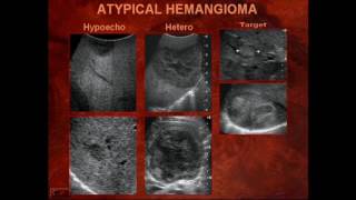
Hemangioma: 3:19
Focal Nodular Hyperplasia: 3:56
Spoke Wheel Appearance: 4:11
Adenoma: 4:28
Hepatocellular Carcinoma: 4:49
Metastases: 5:21
Metastases With Fatty Liver: 6:19
Grade 1 Steatosis: 6:27
Grade 2 Steatosis: 6:56
Grade 3 Steatosis: 7:19
Cirrhosis: 7:35
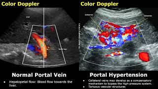
Portal Hypertension: 8:29
Starry Sky Sign (Hepatitis): 9:05
Outro: 9:27
You can support our work with a donation:
Airtm: drsamslibrary@gmail.com
Patreon: patreon.com/drsamsimaginglibrary
Related: longitudinal, sagittal, view, plane, sonography, pictures, scan, imaging, images, probe, transducer, medical, radiology, radiological, ultrasonography, transabdominal, tas, abdominal, pathology, diseases, disease, grading, on, transverse


![CAMPปลิ้น | EP.75[2/2] คุยแบบนี้แล้วจะจบกี่โมงพ่ะย่ะค่ะ!!!](http://i.ytimg.com/vi/N6E8njWH7Zg/mqdefault.jpg)


Watch More Ultrasound Videos: th-cam.com/play/PL4cRFWfjMmf_P02uIGRTFiYNozGKuILAX.html
Abdominal Ultrasound: th-cam.com/play/PL4cRFWfjMmf_8Rxbgv4Ru77mxhAE9mN2L.html
Gynecological Ultrasound: th-cam.com/play/PL4cRFWfjMmf-J4vhXPWZbx5uRIwq77EBd.html
Obstetric Ultrasound: th-cam.com/play/PL4cRFWfjMmf-M-IH-Bq-LY-07LeEFdi5T.html
This is amazing
Dear dr . I can’t thank you enough for all these videos . Doctors like you are blessings for budding radiologists ❤
Thank you very much for watching and Best Of Luck!
Thanks so much for your videos! Passed my ARDMS (OB/GYN) because I watched your videos so much. Now I’m reviewing for the abdomen exam and am once again watching your videos. I really appreciate the time and effort you put into these!
Congratulations on passing your ARDMS (OB/GYN)! Thank you very much and Best Of Luck!
Thank you Dr. Sam, I’ve been a tech for almost 4 years already and learn something new from these videos. It’s a great reference for me. Thankful for sharing your knowledge.
Most Welcome and thank you very much for watching!
I really wanted to thank you for these very informative and well-made videos, you are giving a great contribution to medical knowledge
Thank you very much! Really appreciate it!
Excellent descriptions
Thanks for watching!
Very good lecture and very help full every lecture like this lacture ❤❤❤
Thank you. This very helpful!
Your Welcome!
Thank you! Superb video
Most Welcome and thanks for watching!
Your work is appreciable......
Thank you very much!
thank you so much dr sams for such a great video
Most Welcome and thanks for watching!
We thank you Doctor Sam . for Sharing this talents of yours
Most Welcome and Thanks for watching!
very grateful for this video
Thanks for watching!
Thanks a lot for this useful lecture on US of the liver!
Most Welcome!
Thank you so much for sharing Dr
My Pleasure!
Thank you Dr. Sam..
Most Welcome!
Thank you sir. Plz keep it up.
Thank you very much for your support!
Thank you Dr, you are a blessing 🙌 ❤
Thank you very much for watching!
I have learnt so much watching this video🙏🙏
Thanks For Watching!
Good Dr and good lacture
Thank you very much for watching!
Thank you so much doctor now am competent in applying color Doppler
God bless you doc 🔥🔥💚💚💚🦅🦅
Most Welcome! God bless you too!
Awesome man
Thank you for watching!
Very well explained sir ,your videos are really helpful in enhancing our knowledge ,Thanks for sharing
Most Welcome and thank you very much for watching!
Thank you Dr Sam
Most Welcome!
Thank you so much
Most Welcome!
Thank you Doctor 😊💯
Most Welcome!
You're a genius thank you very much
Most Welcome And Thank you very much for watching!
Thank you sir.
Most Welcome!
Thank you very much. I am learning about my ultrasound and fatty liver.
Most Welcome and thanks for watching!
Thank you Dr. very helpful videos for DMS students too
Most Welcome!
Thank you so much 🖒
Most Welcome!
Helpful ♥️
Thanks for watching!
Very good images 👌
Thank You Sir!
Very good topic
Thanks for watching!
jazakAllah.for ur good work.
Most Welcome!
1:47 HYDATID CYST 2:15 CALCIFIED HYDATID CYST 2:33 HEPATIC ABSCESS
Sir your videos are excellent and superb.
Plz make a video on different inguinal hernia
Thank you very much for watching! Yes there will be a video on hernia in the future.
Amazing
Thanks for watching!
Thank you,
Most Welcome!
u explained very well 👌
Thank you so much!
you are a genius !
Thank you very much for watching!
Thank you
Your Welcome!
Thanx.
Your Welcome!
Nice video
Thank you!
Thank you so much for your explanation Dr. But we wann more about portal hypertation by making it separate topic.
Thanks for watching! Yes, it will be uploaded in the future.
And B Scan..detailed videos on ophthalmic problems..and face ultrasound regarding parotid and salivary glands..all small parts videos...Thank you very much
Thank you for your suggestions! Yes, they will be uploaded in the future. Thanks again!
Many thanks for sharing this video, can i ask you why portal vein margins are iperecoic and other hepatic veins no?Perhaps, becouse the fat blood drained from the bowel?
Most Welcome! Portal veins appear with echogenic borders on ultrasound. This characteristic appearance is attributed to the presence of a dense fibrous tissue sheath that surrounds the portal vein branches. This sheath is also known as Glisson's capsule. This strong reflection at the interface creates the appearance of a bright, echogenic border around the portal veins. Hepatic veins do not have a significant fibrous sheath like portal veins. They are enveloped by a thin layer of connective tissue that does not provide the same level of acoustic impedance as the fibrous sheath around the portal veins.
Many thanks for the explication 🙏
Most Welcome!
Appreciate your efforts ..may you present doppler limp
Thank you very much! There are some videos available but there will be more in the future!
Great
Thanks for watching!
Sir can u make a detailed video on kidneys for medical transcripnist
Thanks for watching! You can watch the video on kidneys. th-cam.com/video/sFdrEwPMzWY/w-d-xo.html
Anatomy of liver please
Thank you for the suggestion!
u rule man
Thanks for watching!
Good afternoon sir i hope you will be fine. If liver appearance is starrysky how to write impression pls reply🙏
Good day! You can write it in this way, "There are multiple scattered bright echogenic foci throughout the liver parenchyma, giving a characteristic "starry sky" appearance."
@@DrSamsImagingLibrary thank u so much sir in impression can write suggestive further investigation or only liver shows starrysky appearance pls reply sir🙏
@@MukeshKumar-gq4ijMost Welcome! It is better to suggest further investigation or write "Clinical correlation is recommended"
@@DrSamsImagingLibrary good night sir ur great 🙏
@@DrSamsImagingLibrary good evening sir i hope you will be fine locular ovarian cyst means sir pls reply🙏
👍👍👍
3:18 HEMANGIOMA 3:56 FOCAL NODULAR HYPERPLASIA 4:29 HEPATIC ADENOMA 4:50 HEPATOCELLULAR CARCINOMA 5:21 LIVER METASTASES 6:28 GRADE 1 FATTY LIVER 6:57 GRADE 2 FATTY LIVER 7:34 CIRRHOSIS LIVER 8:28 PORTAL HYPERTENSION 9:06 VIRAL HEPATITIS
Sir fetus abnormalities video plz sir
Thanks for watching! Yes, stay tuned for more videos!
Sir liver ka normal size kya hota h mm & cm me
Hi! You can watch this video regarding liver measurements. th-cam.com/video/A3akbgBKmcw/w-d-xo.html
Craniocaudal length is 15cm/150mm approximately
@@DrSamsImagingLibrary ok sir mai dekhti hon
@@DrSamsImagingLibrary thanks for your help sir
♥️❤️💓👍💖💕💞🥰
sir, m medical transcriptnist want to earn usg images perfectly!
Best Of Luck!
@@DrSamsImagingLibrary thanks
Report
Thank you so much
Most Welcome!