ไม่สามารถเล่นวิดีโอนี้
ขออภัยในความไม่สะดวก
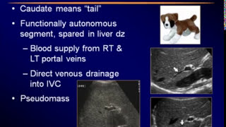
LIVER SEGMENTS & COMMON HEPATIC LESIONS ON ULTRASOUND || DR SANJEEV MANI || LIVER ULTRASOUND TIPS

ฝัง
- เผยแพร่เมื่อ 14 ส.ค. 2024
- This video is brought to you by IndianRadiologist - www.indianradiologist.com
__________________________
Sonobuzz Onsite March 19-20 2022.
register now (limited seats only) pages.razorpay...
This is an On site In Person event with Lectures, Quiz Contests, Discussions, and MMC (state Council points)
_________________________________________________________________________
Follow us on Social Media for Event info, New videos, Free Classifieds Information on Jobs & Machines, Unusual & Rare Radiology Images, New Product Reviews & More
TH-cam: Subscribe & Click on the Bell Icon for notifications: www.youtube.co...
FACEBOOK: / indianradiologist
TH-cam: / indianradiologist
INSTAGRAM: / indianradiologist
Quick learning videos on Radiology for UG and Residents in Radiology. Subscribe to Indian Radiologist and get free Radiology teaching videos from experts in the field of Radiology.
LEARN ABOUT liver abscess, amoebiasis, liver metastatis, cirrhosis, liver tumor, hepatoma, hepato cellular carcinoma, liver lesions, hepatic tumors, usg liver, hemangioma, hepatic cysts
Liver sonography, liver lump, liver lesions, liver cyst, liver tumor, Liver ultrasound, pain on right side


Fabulous explanation of anatomy & a great collection of common cases with perfect segmental localisation… Thanks for another great lecture
L
Lucid ,illustrative and very useful video in our day to day practice ,thank you very much sir
Very nicely explained & presented
Thank you sir, few months ago I requested for this lecture.....
Next time please make a video on EHPVO Vs NCPF
What a simplified,clear explanation of a very important topic of day to day practice!!
Superlative lecture 👌
Keep going @ Indian Radiologist ❤
Extremely helpful ,please Sir conduct more such classes for us. Thanks a lot.
Your speech is very interesting so amazing sir
Very informative
Thank u so very much for making such a difficult concept so easy , for beginners .
Thankyou very much sir
May God bless you sir
AWESOME /-/LUCIDLY EXPLAINED/ILLUSTRATIVE CASES-ALL ROLLED INTO A PAR EXCELLENCE LECTURE-SUPERLATIVE INDIAN RADIOLOGIST
Very much informative
Excellent teaching presentation 👏👏👏👏👏
Excellent explanation about the topic ... Great job .. Thanks a lot sir...🙏🙏
thank you
Very well explained lecture , thank you so much sir
Fabulous thanks Sir
Excellent .
Nice explained
Very nicely explained ,thank you
Very nice clear explanation and plenty of good cases
Thank you Mani sir
So much informative in a simplified way
Thanks a lot
This video strengthening my knowledge Thank you Sir
Excellent explanation. Thanks alot!
Awesome Lecture 👌
Really simplified liver sonography
Great lecture 👌
Tq you sir for giving us good anatomy of liver
😢very needed video Thanks Sir
Nice explanation sir
Very good explain
Nice explain
Awesome simplified explanation sir thank you 🙏
Great lecture ,thank you for sharing
Nicely explained
I really understand it to the fully
Thank you for making a vase topic short
Glad it was helpful!
Very lucid talk.Thank you.
Please do more sessions like this
Great lecture! Very informative and well explained! Thank you
Glad it was helpful!
Excellent presentation
Glad you liked it
Good job 👏 sir
thanks a lot very informative video
Beautiful presentation.
Thank You Sir for simplified lecture
Thanks for liking
Very helpful thank you
Just a little correction "its acoustic enhancement behind a cyst, not shadowing". You used the word "shadowing" twice. Regards
Great explanation sir , Hat's off you
Superb!!!
Great sir
❤️❤️❤️ @indian radiologist
Excellent lecture sirji
Very informative. Thanks.
EXCELLENT TEACHING👋👋👋👋👋💥
Very nicely explained
Votre vidéo est excellente, mais dites moi comment faire pour obtenir une image aussi nette sans interférence de gaz intestinaux !?
Thank You Sir 😊
Very nice lecture
Very clear explanation, thank you Doctor. My ultrasound whole abdomen Impression reads Mild fatty hepatomegaly with hepatic segment VI subcapsular small simple cyst. Kindly advice on how to treat in terms of diet or vitamins to stop any advancement. Is this progressive or something to be worried? Kindly advice. Thank you.
Subcapsular small simple cyst of size 23 x 15mm.
Sir in my opinion just follow up is required...no need to worry about the cyst.
V v informative . thanks
EXCELLENT TEACHING
Would you explain how a hemangioma comes into play? An example would be very helpful.
Thx. GL
Thank you
Well demonstrated
Thank u sir..
Great
Good one
thank you
Awesome... thsnk you
Ultimate explained!!!
thank you🙏🏻
Well explained 🙏
I have cancer in my T6, T10, T11, T12And pelvis. The Dr see that the right side of the liver has lesions. Will it be wise to get have of the liver sugerically removed and chemotherapy? Will oral chemotherapy help or should I get IV type of chemotherapy?
Thank you sir!
They found some lesions on my liver through ultrasound, they want me to do MRI for a better characterization. What chances is this cancer?
AA briliant
Thank you so much, finally i can understand
Glad it helped!
Thanks DR
Thank you sir
Enjoyed🌟🌟🌟🌟🌟
@indian radiologist Dr, what kind of treatment is usually given for comet tail artifact in gallbladder? My ultrasound shows one. Hvng pain in d the lower abdomen. Hvng fibroid too. Im scared to the core and hvnt taken the report to anyone fearing something big.
nothing big, not to worry. consult your gastroenterologist for medical opinion
Dear sir.... Everything you described is true to me but the hydatid cyst you described I think its ball of yarn /wool sign ..... instead of water Lilly sign... Kindly give your feedback..
Hello Krishna. Thank you for watching 🙏. There are many appearances of hydatid but the pic of water lily as shown in this video is quite classical
radiopaedia.org/articles/water-lily-sign-hydatid-cyst?lang=us
Sir can ulcers of colon can be seen in sonography or ct scan?
Colonoscopy is the investigation of choice for the above
@@IndianRadiologistok sir thanks a lot🙏🏻😍😊
Cranial side on which side sir?
Nice
GOOD
😢
Soo good thank u sir