You offer one of the best radiology resource available on the internet. You are my personal favourite. Concise but so much relevant information. Wishing you the best ❤️ Ps- if you could kindly upload content more often. God bless !
I just love the short format videos. The cases are excellent also. I think You really nailed the sweet spot regarding length, yet it is very information dense. Its also great that You repeat the crucial pieces of info multiple times. Definitely one of the best radiology channels.
very useful video from MRI technologist standpoint as we really need to take care of the timing and now I know more clearly about reasons behind it. Thanks
Thanks Solon, I'll keep that in mind! I do show an example of THAD in my hepatic hemangioma part 2 lecture: bit.ly/Hepatic-Hemangioma-2. At minute 05:43, and I talk about THAD/THED (transient hepatic arterial difference/transient hepatic enhancement difference) as it relates to hemangiomas associated with arterial portal shunt, with CT and MRI examples.
Thank you Dr Kowal! Very informative video, though the MR info sounds like Greek to me as a CT tech ;-) I am wondering what your recommendations are for flow rate on this multi-phase liver protocol? As a traveler, standard injection rates vary greatly from facility to facility. For instance, where I am now their standard is 2ml/sec for routine abd/pel and such type of studies. To me, this seems too slow for this particular scan. I would think at least 3ml/sec, if not 4; making the rate closer to an angiogram. Do you find better enhancement using bolus tracking or fixed delay for late arterial phase? If using BT, where do you like to have the techs place the ROI (or visually watch it) and is there a post threshold delay? For instance, if tracking at the aorta near the hepatic artery, do you then wait several seconds to start the scan so that it is enhanced the way you describe? I work mostly in rural facilities and we rarely do any multiphase; with liver being the least performed (renal most often, pancreas second). Thank you for any and all input!
Glad you found it helpful Deana! Typically 2-3 mm/second is required to sufficient enhancement with multiphase liver CT. At my institution, we typically use bolus trigger for all angiograms, but just a fixed delay for late arterial phase.
Excellent. Thanks for ur effort. I just have a question. During late hepatic arterial phase, how does contrast comes into PV? Is it from Aorta to SMA to capillary and then SMV & PV?
Thanks Andy! There are a few CT findings with hepatic abscess that we don't typically see with tumor. The "double target" sign is when we see the enhancing wall of an abscess surrounded by hypodense parenchymal edema. Also, the "cluster" sign of an abscess is when we see multiple small locules giving a clustered appearance. Gas may also occasionally form in liver abscesses as well. Check out this RadioGraphics article for a nice example of these findings: pubs.rsna.org/doi/full/10.1148/rg.2016150196
Hi Liz. I’m in same situation. They found lesions in my liver from ultrasound and want me to do MRI for further investigation but I don’t want contrast. Any updates on yours?
@@LVH100 Hi Tony, she wouldn't do the MRI but just had another ultrasound and was thought to be hemangioma which is usually found incidentally on ultrasounds.
@@Radquarters Thank you . It's great . I have been struggling for year to find a method for reading those with confidence , I always have the feeling that I am misssing something !
With the late arterial phase (where hepatic veins are not opacified) how then can we differentiate the liver segments accurately for anatomical resection if there is already washout in PV phase?
While certain liver masses show washout in the PV phase, the hepatic and portal veins will be well opacified during this phase and can be used as landmarks to determine the anatomic segments.
Hello , The HQ of radiology . Please can you give me more precisions about Multiphasique Hepatic CT scan ? quantity of contrast ? and rate (Q) of injection ? what is best , bollus traquing or fixed delay ? Thanks a lot . We missssssssss you !!!!!
I scrolled through the comments looking for this exact info! It is one of the few that were unfortunately not answered 😕 I mainly need/want to know his recommendations about rate of injection, as it makes a difference to when the contrast will reach the specific phases. Also curious about bolus tracking, where to place roi (or visually watch it) and if there should be a post threshold delay. I am going to try and post my own comment and see if he answers.
i don't quite understand why u mentiond that hypervascular tumors get most of the blood from the hepatic artery.not portal vein? could y help me explain that,thanks a million
Although liver parenchyma is fed by both the hepatic artery and portal vein, hypervascular tumors such as hepatocellular carcinoma (HCC) may be fed primarily by the hepatic artery, and that is why they appear hypervascular. With HCC, angiogenesis can also occur, which is the creation of new blood vessels. This phenomenon leads to increased arterial flow and a gradual reduction of portal venous flow to the mass. It's not a hard and fast rule though, as the arterial supply to HCC has been shown to vary based on tumor grade.
The portal venous phase varies depending on using a fixed time delay (usually 60-90 seconds) versus bolus tracking (50-60 seconds), but otherwise looks similar on CT vs. MRI.
@@jeevnasam4810 You might want to consider a delay closer to 35 seconds for the late hepatic arterial phase. 7-8 seconds is typically closer to a true arterial phase (i.e., hepatic artery enhancement with no portal venous enhancement).
patients might have different hyper or hypodinamic CO, do you adjust your timing to that . Do you note a differente in the timing of the arterial or portal phase between child A patients that to Child C patients because of Different cardiac Output ?
Good question Luis! When we use a fixed (AKA standard or empiric) contrast timing delay, we definitely see differences in the appearance of the immediate post-contrast phase depending on the patient's cardiac output. One way to prevent this variation is to use a trigger to delay for contrast using bolus tracking, where we sample the liver or aorta with an ROI at timed intervals after injection of a contrast test bolus, and then when the density reaches a certain threshold, the start of the scan is triggered. This technique give a more consistent, uniform pattern of contrast enhancement for each patient, but comes at the cost of a slightly increased radiation dose for the patient, technologist time, and increased contrast volume. Hope that helps!


wow ! Your presentation is amazing and smart. Very grateful of you. Thank you 🙏
Thank you, that's very kind! You're most welcome
Great and illuminating presentation , Dr Daniel!
Thank you, and so glad you enjoyed it!
Wow, excellent, concise and very clear lecture for surgeons! Thank you very much 👍
Awesome, great to hear that Aldwin Ong, thank you!
These are gems that are great for initial study and short refreshers. Top notch
I'm thrilled to hear that Peregrine, thank you!
You offer one of the best radiology resource available on the internet. You are my personal favourite. Concise but so much relevant information. Wishing you the best ❤️
Ps- if you could kindly upload content more often. God bless !
Wow, thank you for the kind words Dr. Fawaz Yousuf! Stay tuned :)
Best lecture I have heard.
Thank you so much!
Amazing video!
Thanks Talia Cheng, appreciate that!
I just love the short format videos. The cases are excellent also. I think You really nailed the sweet spot regarding length, yet it is very information dense. Its also great that You repeat the crucial pieces of info multiple times.
Definitely one of the best radiology channels.
Awesome, thank you Tenzin Angio! Really appreciate that. I try to keep the videos short and high-yield, since time is our greatest commodity :)
Informative lecture. Useful for us CT Techs. Thanks.
You’re welcome, glad you found it helpful!
This is perfect for beginner radiologists. Thank you for your work 🙏
Glad it was helpful Hammer Radiology!
Great lecture
Glad you think so!
Great to learn some abdominal radiology knowledge. Great teacher.
Thank you kindly!
Excellent lecture ! Thanks.
Very Nice explanation. Thankyou
You're welcome, and I'm glad you enjoyed it!
Excellent presentation !!! many thanks
That was an awesome lecture.....Thanks about
Most welcome!
Thank you so so much for making such a great video! As an intraining newbie, this helps me a lot. Thank you!
Awesome, glad to hear that Ramita!
That was a great presentation
Thanks so much Ashok!
OH, EXCELLENT LECTURE SIR, MUST APPRECIABLE.
The best one I have ever seen. Soooo easy to remember. Thanks!
Glad you liked it XING Xing!
Thank you for helpful lecture 🙂
Most welcome!
Great video ❤
Glad you liked it!
Very lucid presentation, thanks
Glad you liked it!
Great video because it's simple and yet, effective! Thx mate.
I try to keep the lectures simple and to the point, so that’s great to hear. Although tangents can be fun too ;) Thank you!
very useful video from MRI technologist standpoint as we really need to take care of the timing and now I know more clearly about reasons behind it. Thanks
Glad it was helpful!
Very nice lecture
Thank you for watching suvarna latha Penukonda!
Excellent video, this is the second time after a few months to review again. will come back again. Thanks a lot.Already subscribed.
Great to hear you found it useful enough to watch more than once!
excellent presentation
Thank you!
Super.thank you
You're welcome, glad you enjoyed it!
Your lecture is awesome. Thank u
Thank you RASHID MAHMOOD, glad you liked it!
This video is very helpful. Thank you.Please do more liver and biliary track imaging.
Thank you! I'll work on that :)
The best video thank you
Thank you, greatly appreciated!
Thankyou so much! Amazing lecture.
You're most welcome Apoorva, and thanks for watching!
Excellent.
Brilliant presentation! Thanks a lot!!!
You're very welcome Ivan, and thank you!
Very nice 👌 thanks
Most welcome 😊
Nice presentation
Glad you liked it Rajesh, thanks!
nicely explained,thanks
You’re welcome!
a very nice video
Than you for the precious lecture!
You are welcome Jiyang Kim, glad you liked it!
extremely helpful. Thank you.
Glad to hear it, thank you KC Okoro!
Very good
Thank you Anjali!
It helps a lot
Great to hear!
great talk
Excellent.Please do a video on THAD
Thanks Solon, I'll keep that in mind! I do show an example of THAD in my hepatic hemangioma part 2 lecture: bit.ly/Hepatic-Hemangioma-2. At minute 05:43, and I talk about THAD/THED (transient hepatic arterial difference/transient hepatic enhancement difference) as it relates to hemangiomas associated with arterial portal shunt, with CT and MRI examples.
Great
Comprehensive
Consice
Thanx
Glad it was helpful!
Great video thanks so much!
I want to ask, when you talk about contrast media you mean the general MRI Contrast i.e Prohance or the liver specific i.e. Primovist?
Glad you enjoyed it!
Amazing
Thank you!
Ooh perfekt . Thank you very much
You're welcome 😊
Well explained. Thanks for your help🇨🇩
Glad it was helpful!
Awesome!
Thank you! Cheers!
Thank you Dr Kowal! Very informative video, though the MR info sounds like Greek to me as a CT tech ;-) I am wondering what your recommendations are for flow rate on this multi-phase liver protocol? As a traveler, standard injection rates vary greatly from facility to facility. For instance, where I am now their standard is 2ml/sec for routine abd/pel and such type of studies. To me, this seems too slow for this particular scan. I would think at least 3ml/sec, if not 4; making the rate closer to an angiogram. Do you find better enhancement using bolus tracking or fixed delay for late arterial phase? If using BT, where do you like to have the techs place the ROI (or visually watch it) and is there a post threshold delay? For instance, if tracking at the aorta near the hepatic artery, do you then wait several seconds to start the scan so that it is enhanced the way you describe? I work mostly in rural facilities and we rarely do any multiphase; with liver being the least performed (renal most often, pancreas second). Thank you for any and all input!
Glad you found it helpful Deana! Typically 2-3 mm/second is required to sufficient enhancement with multiphase liver CT. At my institution, we typically use bolus trigger for all angiograms, but just a fixed delay for late arterial phase.
Excellent.
Thanks for ur effort.
I just have a question.
During late hepatic arterial phase, how does contrast comes into PV? Is it from Aorta to SMA to capillary and then SMV & PV?
From splenic vein
Thank you
You're welcome
just beautifully done merci bcp
Hi thanks for the nice presentation!
Can I ask If theres any challenge to distinguish between tumor and abscess @ liver on CT conclusively ?
Thanks Andy! There are a few CT findings with hepatic abscess that we don't typically see with tumor. The "double target" sign is when we see the enhancing wall of an abscess surrounded by hypodense parenchymal edema. Also, the "cluster" sign of an abscess is when we see multiple small locules giving a clustered appearance. Gas may also occasionally form in liver abscesses as well. Check out this RadioGraphics article for a nice example of these findings: pubs.rsna.org/doi/full/10.1148/rg.2016150196
What if you are someone who is getting an MRI on the liver because of lesions found but don't want to take the contrast. Can the MRI still be done?
Hi Liz. I’m in same situation. They found lesions in my liver from ultrasound and want me to do MRI for further investigation but I don’t want contrast. Any updates on yours?
@@LVH100 Hi Tony, she wouldn't do the MRI but just had another ultrasound and was thought to be hemangioma which is usually found incidentally on ultrasounds.
Please do a videos on your search pattern on body mri .
Hi, I do plan on doing that type of video as part of a "How to Read" series in the near future. Thanks for the suggestion!
@@Radquarters Thank you . It's great . I have been struggling for year to find a method for reading those with confidence , I always have the feeling that I am misssing something !
great, great, great
Thank you Frank Robert, glad you enjoyed it!
da best!
Nice, thanks Vincent!
Pure gold!
Great to hear, thank you Dean!
Great
Thank you!
Thank you so much pro
You are welcome!
With the late arterial phase (where hepatic veins are not opacified) how then can we differentiate the liver segments accurately for anatomical resection if there is already washout in PV phase?
While certain liver masses show washout in the PV phase, the hepatic and portal veins will be well opacified during this phase and can be used as landmarks to determine the anatomic segments.
Hello , The HQ of radiology . Please can you give me more precisions about Multiphasique Hepatic CT scan ? quantity of contrast ? and rate (Q) of injection ? what is best , bollus traquing or fixed delay ? Thanks a lot . We missssssssss you !!!!!
I scrolled through the comments looking for this exact info! It is one of the few that were unfortunately not answered 😕 I mainly need/want to know his recommendations about rate of injection, as it makes a difference to when the contrast will reach the specific phases. Also curious about bolus tracking, where to place roi (or visually watch it) and if there should be a post threshold delay. I am going to try and post my own comment and see if he answers.
Hello sir, how much volume of contrast and flow rate should be followed in hepatic protocol
Hi, it varies depending on institution, but typically we give 100 cc Omnipaque 300 at 4 cc/sec.
Subscribed sir
i don't quite understand why u mentiond that hypervascular tumors get most of the blood from the hepatic artery.not portal vein?
could y help me explain that,thanks a million
Although liver parenchyma is fed by both the hepatic artery and portal vein, hypervascular tumors such as hepatocellular carcinoma (HCC) may be fed primarily by the hepatic artery, and that is why they appear hypervascular. With HCC, angiogenesis can also occur, which is the creation of new blood vessels. This phenomenon leads to increased arterial flow and a gradual reduction of portal venous flow to the mass. It's not a hard and fast rule though, as the arterial supply to HCC has been shown to vary based on tumor grade.
Thanks
You're welcome!
That was neat!
Thank you!
Why the portal venous phase timing in ct is 80 sec and in mri 40?
The portal venous phase varies depending on using a fixed time delay (usually 60-90 seconds) versus bolus tracking (50-60 seconds), but otherwise looks similar on CT vs. MRI.
Super!
Thank you!
নাইস বন্ধু
Thanks Prosanta!
The Best
Thank you!
Can we do it in ge 16 slice machine.. we don't get this kind of scan ..our del ded is 8
Yes, a multiphase liver scan can be performed with a 16 slice scanner.
@@Radquarters yes sir but we r not getting proper arterial phase our scan del sec is 7 to 8 sec
@@jeevnasam4810 You might want to consider a delay closer to 35 seconds for the late hepatic arterial phase. 7-8 seconds is typically closer to a true arterial phase (i.e., hepatic artery enhancement with no portal venous enhancement).
patients might have different hyper or hypodinamic CO, do you adjust your timing to that . Do you note a differente in the timing of the arterial or portal phase between child A patients that to Child C patients because of Different cardiac Output ?
Good question Luis! When we use a fixed (AKA standard or empiric) contrast timing delay, we definitely see differences in the appearance of the immediate post-contrast phase depending on the patient's cardiac output. One way to prevent this variation is to use a trigger to delay for contrast using bolus tracking, where we sample the liver or aorta with an ROI at timed intervals after injection of a contrast test bolus, and then when the density reaches a certain threshold, the start of the scan is triggered. This technique give a more consistent, uniform pattern of contrast enhancement for each patient, but comes at the cost of a slightly increased radiation dose for the patient, technologist time, and increased contrast volume. Hope that helps!
@@Radquarters thanks a lot
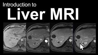
Those first hypOvascular liver masses looked like cysts 😅
They do a look a bit deceptive, but their density measurement was above fluid. Thanks for watching!
5:35 delay phase
👍👍👍👍👍👏👏👏👏👏👏👏
Thanks!
🙏🏼🙏🏼🙏🏼
Thanks for watching Nagi!
Very nice
Thank you!