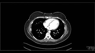
Interpreting CT Abdo: Background Liver, Hepatic Steatosis, High Attenuation Liver

ฝัง
- เผยแพร่เมื่อ 8 ธ.ค. 2021
- Access our CT and MRI case-based courses at navigatingradiology.com, which include fully scrollable CT abdomen and pelvis cases, walkthroughs of imaging findings, and comprehensive reviews of basic and more advanced imaging studies.
Intro to looking at background liver 0:22
Hepatic steatosis (fatty liver) 2:40
High attenuation background liver 8:10


Thank you for the high-quality video. It was on point, with a clear voice, a good explanation, and CT images. Keep up the good work. 😀
he's back!
Your series are so helpful. Welcome for your high quality tutorials.
Glad you're back!! Great video as always.
Super video. Looking forward to the next videos. Great to have you back.
smooth and extremely clear presentation! Congrats & thanks for sharing
Thanks. Your tutorials are really helpful!
Was Waiting for you!
You made it easy for us sir. Thank you!
0:19 FOUR PHASE SCAN OF NORMAL LIVER 3:19 CT DENSITY OF A NORMAL LIVER 4:16 MODERATE TO SEVERE STEATOSIS 8:32 HIGH ATTENUATION LIVER
Excellent learning source! Thanks heaps!
Excellent video
Welcome back!!
Thanks a lot
Thanks very much 🥰🥰🥰
Thank you so much pro
Routine CT moderate to severe fatty liver, compared to the spleen. hounds per unit.
I want to ask. A few days ago I did a Contrast CT Scan of the Abdomen and Pelvis. The doctor didn't tell me to change my pants and shirt because he saw that I wasn't wearing any metal. but I forgot to tell the doctor that my underwear has plastic buttons. will this cause artifacts
No issue
Waiting for the cirrhosis part
I had a liver scan and it’s like dude how the heck can they really tell what’s going on! The doctor couldn’t tell if a nodule was benign or malignant and if my body isn’t hurting then why would I care or even want to know ?! Some of these tests as doctors like to do are in conclusive and unnecessary and unreliable and a inconsistent diagnosis and inconclusive inconvenience 😂
If they do a routine scan looking for something acute (and not specifically to look for and characterize liver lesions), they may see something in the liver that is indeterminate on that limited scan. Dedicated liver imaging will characterize it much better in most cases. That’s just how it goes - tests are designed for a specific purpose and if they pick up something incidentally then you need to rely on the clinical history and/or more imaging. Unfortunately there’s no magic single test for everything