POCUS - Lower Extremity Deep Venous Thrombosis (DVT) - Updated

ฝัง
- เผยแพร่เมื่อ 30 พ.ค. 2024
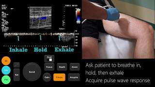
- Update video of DVT ultrasound -- In this video the basic anatomy and technique for point of care lower extremity DVT ultrasound is discussed.
- วิทยาศาสตร์และเทคโนโลยี


Great video, but I would have loved to have seen an example of a thrombus formation.
Thank you for this fantastic video, extremely useful!
Thank you for making this understandable! I wish you’d make a calf vein video for those of us who have to go all the way down the leg for DVTs. 🙂
Probably the best video on DVT ultrasound.Thanks!
Glad it was helpful!
I disagree. Skipping and not checking lower into the calf is unacceptable.
Very nice practice I really appreciate this Chanel
Hi Marx, thank you so much for the informative and detailed video. Could you upload a video for Femoral Duplex and Carotids, please? I really appreciate it, thanks.
Very useful presentation 👏👏👏👏👏
Thanks Jared. Nice video and it helped me with just in time education
Very illustrative video and excellent explanation 100% satisfaction
Glad you liked it!
Excellent teaching
Sometimes we need anterior/posterior tibial vein puncture due to throbosis within popliteal vein. Looking forward another video to show tibial vein puncture. Thank you !
Thank you Sr your videos are very helpful for well explained understandable.
Thanks for sharing your knowledge
Wow!!! TH-cam are great+the best! Even better than real ultrasound lessions. Thank you soooo much. You win one more fan!
Please do another video from distal leg also.
Best greetings from Germany :))
Unfortunately I don't scan the distal (calf) veins so would not be the best resource for this. If you are a physician performing point of care ultrasound there are not any studies I'm aware of validating physicians looking at the distal veins - if you are a vascular sonographer this would be typical for that to be in your skill set and taught.
The sensitivity, however, of ultrasound finding a distal calf vein is poor and many feel that it should not be done. Additionally there is much controversy regarding treating distal vein DVTs. Several studies have shown that the risks of bleeding due to anticoagulation are much higher than the benefit.
Hope that helps. If you need any resources/articles regarding that feel free to email me and I can send them to you.
@@POCUSGeek thank you so much for your feedback. Please let me know the names of this articles-study results from the distal leg ultrasound, so I can argument better in the clinic . Best regards.
Remember when discussing this that everyone has different experiences and has a different threshold for treatment . The problem with dvt in general is that it can lead to significant morbidity, however, this generally relates to proximal dvt but we often extrapolate that morbidity to calf DVT.
Distal (calf) DVT has not be studied as extensively as proximal DVT so there is still much dispute on what to do.CHEST guidelines do say a negative whole leg ultrasound effectively rules out any DVT and no follow up is needed. The issue with that is that the sensitivity for whole leg ultrasound is only about 70-75% for isolated calf dvt. With a poor sensitivity, in my opinion, this does not rule out a DVT of the calf veins and proximal extension could still occur.In fact, CHEST guidelines recommend D-dimer and proximal leg ultrasound as first line tests for the different risk groups - low, moderate and high risk.
For moderate and high risk patients with negative proximal DVT ultrasound but positive d-dimer testing they recommend repeat proximal leg ultrasound in 1 week.
The suggested workup gets a little long so you should look at the documents linked below. I'll try to do a video sometime in the near future to address these points.NICE guidelines are similar to the CHEST guidlines.You can find both here:
journal.chestnet.org/article/S0012-3692(12)60128-7/fulltext
journal.chestnet.org/article/S0012-3692(15)00335-9/fulltext
www.nice.org.uk/guidance/cg144/resources/venous-thromboembolic-diseases-diagnosis-management-and-thrombophilia-testing-pdf-35109570835141As far as treating calf DVT; here a number of articles that have contradicting opinions:Robert-Ebadi, H. and M. Righini, Should we diagnose and treat distal deep vein thrombosis?Hematology Am Soc Hematol Educ Program, 2017. 2017(1): p. 231-236.
Woo, K.M. and J.K. Goertz, Diagnosis And Management Of Deep Venous Thrombosis In The Emergency Department.Emerg Med Pract, 2015. 17(3): p. 1-24; quiz 25.
Stone, J., et al., Deep vein thrombosis: pathogenesis, diagnosis, and medical management.Cardiovasc Diagn Ther, 2017. 7(Suppl 3): p. S276-S284.
Palareti, G., Do isolated calf deep vein thrombosis need anticoagulant treatment?J Thorac Dis, 2016. 8(12): p. E1691-E1693.
Kearon, C., et al., Antithrombotic Therapy for VTE Disease: CHEST Guideline and Expert Panel Report.Chest, 2016. 149(2): p. 315-352.
Kitchen, L., et al., Emergency Department Management of Suspected Calf-Vein Deep Venous Thrombosis: A Diagnostic Algorithm.West J Emerg Med, 2016. 17(4): p. 384-90.
Righini, M., Is it worth diagnosing and treating distal deep vein thrombosis? No.J Thromb Haemost, 2007. 5 Suppl 1: p. 55-9.
Utter, G.H., et al., Therapeutic Anticoagulation for Isolated Calf Deep Vein Thrombosis.JAMA Surg, 2016. 151(9): p. e161770.
@@POCUSGeek wow! Thanks 1000 times for all your explanation+time! :)). *great* !
Very nice understandable video
Thanks so much . I am not familiar with vascular ultrasound . Your video does hep a lot . Please make more more videos for the beginners and advance sonography tech in vascular
Commendable and v helpful.
I just got a ultrasound done from the back of my knee to the groin like the one in this video. I went to my local emergency department with swelling and pain in my calf yesterday. Today I went back for the ultrasound. My question is if the pain and swelling is in the calf how come that area wasn't scanned ? The nurse and the tech who performed the ultrasound told me that they don't do the whole leg when checking for DVT. I was just confused by this and was looking for further clarification. If you could provide reasoning as to why they don't scan the whole.leg that would be a huge help to ease my mind ! I'm not looking for specific medical advice. It's just you said you don't scan the lower let either so I figured you may be able to provide more insight. Thanks in advance !
Thanks ,good information
super good! 👍
Thank you so much, absolutely great video. Please let me ask a question about the diagnostics of DVT in pregnant patients: what are ultrasound criteria to exclude obstructions of the pelvic veins? Thx so much
Thank you so much
Very educative thanks 🙏🏿 a lot
Thanks.. It is very fine🌺
Are you planning to speak about DVT in the upper extremities?
I hope to eventually when time permits
Or DVT in the pelvic, abdominal, or thoracic cavities?
Thanks a lot for the nice video, you made this very simple, as you go step by step,
I NEED HELP DIFFERNTIATING PTVS AND PEROS AT THE POP TRIFURCATION. CAN YOU DO A VIDEO OR POST LABELED IMAGES PLEASE?
I’ve just been diagnosed with “extensive venous thrombus extends from the adductor hiatus level dismally, no more proximal involvement”. My GP seemed I’ll equipped to explain. I want to know 1) where exactly does the clot begin and end; 2) what vein/artery is blocked; 3) what exactly occurs when the vein “dies”? Which is what my GP said will happen. Varicose veins? Where? What else happens down the track? Can you answer any?
what if you compress and the vein collapses half way?how do you know its a clot and not your strength of applying the pressure?especially when the leg is edematous
Hi...the video is very informative. It was discovered I have a small blog clot on my lower calf. My Dr put me on xarelto...also I have a inflammation behind the knee...ultrasound revealed it was not cyst but no exclamation was given to me On what it might be...any thoughts would be much appreciated. Thanks
hi! ive been told that when it comes to VENOUS doppler exams, there is NO NEED to ANGLE CORRECT the cursor parallel to the vessel, but i still see some techs doing it. would you mind clarifying this for me?
Angle correction gives a better spectral tracing and thus makes the interpretation easier. Otherwise, there’s no need for angle correction.
Thank you sir,can you please down load a video for longgituninal view for lower extremity vein duplex scan with valsalva maneuver thanks sir.
May you help us with other vedios on arterial lower limb system
Thank you please upload video providing guidance on report writing for doppler ultrasound with findings
Here are some reporting guidelines provided by American College of Emergency Physicians (ACEP) - www.acep.org/globalassets/uploads/uploaded-files/acep/clinical-and-practice-management/policy-statements/information-papers/emergency-ultrasound-standard-reporting-guidelines---2018.pdf
Both my lower legs are very red swollen very painful .I have been having clot injections in my stomach for 6 days now while I wait for a scan my legs are killing me
Perhaps things have changed, but my understanding is that ultrasound is a screening technique, while venography is the gold standard for clinical diagnosis.
You are correct that venography is considered the gold standard but almost never used. US is more readily available and still has great sensitivity/specificity for proximal leg lower extremity DVT especially when serial exams are used.
www.ncbi.nlm.nih.gov/pmc/articles/PMC3278049/ - chest guidelines
A lot of publications are finding that A large persentage of PEs are originating from the Gastrocs, perineal veins. These should always be visualized.
Thanks for the comment. This is an interesting topic and can lead to a lot of discussion. Could you share some of those articles here?
Previous guidelines for CHEST and NICE have only recommended proximal leg ultrasound which is to the popliteal vein. It's my understanding this is Wells' Score is based solely off of proximal leg ultrasound. Here's a link on NICE's recent recommendations www.nice.org.uk/guidance/ng158/resources/visual-summary-pdf-8709091453
Whole leg ultrasound that you refer to may be utilized in practices but there is much debate in whom you would treat with anticoagulation if a debate is found. Risk vs benefit but be weighed. Additionally whole leg ultrasound hasn't had great sensitivities which would not rule out a DVT.
Using D-dimer and proximal leg DVT is the basis of the recommendations with repeat ultrasound in certain individuals. In this playlist is a simplified breakdown of CHEST guidelines for those that are using point of care ultrasound. You'll have to skip to videos 3-5. th-cam.com/play/PLMoMZ6-wVYgdwcfTVJ59s9CsS-Pk9MukA.html
My name is akinola and i am certified in vascular technology. Do you teach Echo? If yes i would like to enroll. I find this video very useful
Resul?
Im trying to watch this and work at the same time so my question may have been answered in the vid but here it is anyway: How important is it to scan and identify the profunda both distal and proximally? tks for the videos. I've been doing u/s for 15 years but just now began doing vascular in the past year. It's not a easy as it looks ppl! lol
Great question. Studies have shown sensitivities of two point compression which involves the saphenous junction and popliteal vein around 95%.
Another study has show that approximately 8% of proximal leg DVTs where located in the femoral vein between these two points and not present at either of the saphenous junction or the popliteal vein. This would include the deep femoral vein junction.
I'm unaware of any protocols that follow the deep femoral vein itself but if you are to visualize it as you follow the femoral vein distally you should make sure the deep femoral vein is compressible also.
@@POCUSGeek Thank you for responding. I hope you continue to make vids. Would love to see real time of a difficult leg if that is possible at all. Sometimes it's nearly impossible to visualize what we need due to pt condition and body habitus and I wish I knew some techniques or 'tricks' to help with that
@@jencrochetingradtech5913 Not sure what options you have as far as machines but for obese patients I chose a linear probe with a lower frequency. 10MHz vs 13 MHz. Also each probe has a frequency range. You could try a lower frequency. You should get more depth with this.
If that doesn't work then I will use a curvilinear probe instead. you loose a little resolution but get the depth you need. In obese patients this often leads to an overall better image.
Hopefully in the future I can put out a video showing the curvilinear probe.
Hope that helps.
Vascular is the devil!
Can a thrombus still exist with compressibility of the vein?
If a vein is completely compressible then there is not a clot. In order to demonstrate that it is completely compressible the vessel walls should be able to touch each other.
A mistake that can happen though is that a clot could be present and with compression the operate interprets the wall touching the clot as complete compressibility.
Hi,I need to have a certificate how can you help
Very clear and useful introduction, Thanks a lot!
💞
What difference between Doppler ultrasound and angiography in diagnosis of DVT we know Doppler it is visualization the blood flow and see sit of thrombosis wherever the anioraphay the use in the lower extremity bescuse Doppler difficult in small blood vessels
I need explain in this information please add your Notes thanks
Sorry arteriography
Venography in DVT in vein arteriography arterial
Are all DVT US done in transverse?
Yes compression should only be done in a transverse view.
There are some protocols that do include long axis views but these typically include use of color and or vascular doppler. This is what will often be used by vascular and/or radiology departments and are referred to as duplex scan. Not typical for point of career ultrasound.
❤😎
Do you answer emails??
I have something I think a doctor missed and just need to know so I can save my life I have a picture can I email you that shows a worm in my leg and we’ll I think its been my problem please get back to me
I'm sorry to hear you are having issues. This is an educational channel only and I don't offer medical advice. Any medical advice should come from your physician/primary care clinician or you could seek a second opinion from another clinician in your area.
@@POCUSGeek yeah I have figured it out it’s schistosomiasis and it’s killing lots of people silently I have found that I am getting exposed by my water so this isn’t good because this is the silent killer fiber clots that everyone was blaming on covid so hopefully this comes to surface so that people can get treatment I can’t believe I find this instead of our science teams and bio team like water company didn’t even catch them why
Did you get the clot shots?
@@kdlittlehawk I had a blood analysis I didn’t have the covid spike or the ring so it’s schistisomoasis
Dismally = distally