Echocardiography Standard Protocol | Step by Step | Complete Trans-thoracic Normal Echocardiogram

ฝัง
- เผยแพร่เมื่อ 20 ก.ย. 2024
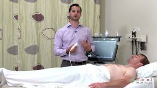
- In this video I am going to illustrate the protocol for performing complete and comprehensive transthoracic echocardiography, which meets the current standards of American Society of Echocardiography. I will not be going into a lot of pathological issues here so the protocol presented here is meant as a guideline and does not cover every aspect, such as off axis views. I am also going to talk basic probe position and major structures visible side by side with the protocol steps.
So when you follow the protocol you don't skip anything, you can get additional images according to the pathology found. I personally found that once I could execute the standard protocol flawlessly, I was able to add and refine additional echo scanning skills while deepening my understanding of the purpose of each echo image. So it's immensely useful to learn basic steps of protocol. We will start with parasternal long axis window followed by short axis, apical and in the end subcostal and suprasternal windows.
👉 Subscribe for more Videos
👉 Share this video
👉 Watch other educational videos:
✅ WATCH Trans-Esophageal Echo TEE Protocol Here: • 16 Views to Master in ...
SHARPEN YOUR SKILLS BY MOCK EXAMS COLLECTION:
✅ ECHO MOCK Exam Part 1: Collection of Thirty Echo Cases: • Echocardiography Mock ...
✅ ECHO MOCK Exam Part 2: • Echocardiography Mock ...
✅ ECHO MOCK Exam Part 3: • Echocardiography Mock ...
✅ WATCH: Collection M Mode Echocardiography Cases: • M Mode Echocardiograph...
✅ HOW TO DO ECHO - COMPLETE GUIDE: www.drmusmanja...
✅ BROWSE FREE ECHO LIBRARY HERE: www.drmusmanja...
Echocardiography protocol Steps | Normal Echocardiogram | Learn How to do echo in 10 min
Following are the key steps according to standard protocol:
00:41 Parasternal Long Axis (PLAX):
2D image with increased depth
2D image with decreased depth
Color Doppler on AV, MV, IVS
M- Mode on Left ventricular level, MV and Aortic root
Measure IVS, LVID and PWT in diastole and systole at MV tip level
Measure Proximal RVOT
Zoom LVOT, measure LVOT diameter, AV annulus
Zoom, color Doppler on Aortic Root, measure at Sinus of Valsalva, SinoTubular Junction and at ascending aorta level
LA diameter in 2D image
01:31 RV Inflow View:
2D Image
Color on TV
CW on TV
02:00 RV outflow View:
2D Image
Color on pulmonary valve
CW on Pulmonary Valve
02:55 Parasternal Short Axis (PSAX)
At the level of great vessels
03:06 At Aortic Valve Level
2D image
Zoom/Focus on TV
Color on TV
CW on TV
Zoom/Focus on PV
Color on PV
CW on PV
Zoom image of LA Appendage
03:50 At Mitral Valve Level
2D Image
Color on MV
At Papillary Muscle Level
2D image
04:48 Apical Four Chamber View:
2D Image
Zoom at Ventricular level
Zoom/Focus on MV
Color on MV
CW on MV
PW at MV tips
TDI Septal and Lateral Mitral annulus
Color on Pulmonary veins
PW on Pulmonary veins
LA Volume in end-Systole
RV Focused View in 2D
Color on TV
CW on TV
RA Area and RA Volume in end systole
TDI of lateral annulus of TV for S'
M Mode of lateral annulus of TV for TAPSE
RV Area in end diastole and end systole for FAC
RV dimensions in end diastole (At Base, mid and mid annulus to apical level)
Apical Three Chamber View:
2D Image
Color on LVOT and AV
CW on AV for IVRT
PW in LVOT
Apical Two Chamber View:
2D Image
Color on MV
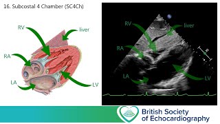
Subcostal View: (SC)
Four chamber view in 2D
Color on IAS
PW on IAS
IVC and Hepatic Veins in 2D and Color Doppler
PW Doppler on Hepatic Veins
Measure IVC Diameter in end diastole
Measure IVC collapsibility index
2D image of Abdominal Aorta
Color on Abdominal Aorta
PW Doppler on Abdominal Aorta
Supraternal View:
2D Image
Color on Ascending Aorta
PW on Ascending Aorta
Color on Descending Aorta
PW on Descending Aorta
CW on Descending aorta
#Echo #Echocardiography #Drmusmanjaved
#cardiology #heart


♦ECHO MOCK Exam Part 4♦Collection of Echo Cases for practice:
th-cam.com/video/oKWBeE1dHmM/w-d-xo.html
♦Echocardiography MCQs: th-cam.com/play/PLdLUoX4jbn3h6nRzhPlLcNWl1kOfQyV9a.html
♦View All Echocardiography MOCK Exams: th-cam.com/play/PLdLUoX4jbn3jFrQJXoBpOSGdnPQzvHh4o.html
thanks alot
I am so grateful to be able to do echocardiograms for a living ❤
Thats Great ♥
I will be having this test tomorrow !!! Pray all will be normal !!! I am 70 years old.
Hope all went normal, Best wishes for you
Best wishes. I had mine. Basically ok with a mitral valve being degenerative but not linking or hard. Good luck Sister.
Recording steps of protocol in a sequence is always useful...excellent
Glad it was helpful!
To be honest cardiology is the only thing that helped me keep having faith in my career path
Great
Best of luck
Great❤
❤
Omg this is a life saver, I look how detailed it is, I would love if you could upload a updated one with how you do some of these measurements, that would be helpful for me since am not in the lab much
Sure! will try to upload soon with the methods of taking these measurements inshAllah
Thanks a lot for great information and details
@@DrMUsmanJaved please, share the link, if you have done a video taking the measurements , will be much appreciated !
Yes sure, i will make a video on it 👍
@@DrMUsmanJavedyes please
So clear and helpful..thanks alot for uploading such useful content..in our lab we prefer to have LV on right hand side.
Most welcome 😊
Worth watching again and again for reference 👌
Thanks
This is beautiful. With speed slowed down to 0.75 and captions on, I have basically 6 weeks of lab missed due to 11 students on 4 machines.
Thanks alot 👍
Jazakh'Allah for this video brother
JazakALLAH brother ♥
Brilliant illustration of all the steps 👍👍👍
Thanks
It is a very clear and useful presentation with great illustrations! Excellent work! Thank you!
Most welcome ♥
EXCELLENT description of basic protocol to be followed in all patients..thanks for sharing
Glad it was helpful! Thanks
Dedicated approach and great job...hope you will upload a content about normal, mild, moderate and severe ranges of measurement of all dimensions and volumes...👍
Thanks ..i will upload soon inshAllah
One of the best detailed vedio I have watched ❤
Glad you liked it ..Thanks Alot ♥ Access whlole Echo library here: www.drmusmanjaved.com/p/echocardiography_23.html
Was looking for this exactly!
Excellent and to the point steps.. Thanks for sharing good with us ❤
Thanks
Definitely saving this video! Thanks a bunch!
Welcome ☺
Thank you so much for this lecture, need more on echo. Wonderful!
Thanks alot ♥ will upload more inshAllah
Very Helpful..Thanks alot for sharing 👍
Excellent description. Thank you ❤
Most welcome ♥ ♥ ♥ ECHO LIBRARY - Watch Echo Cases, Tutorials, MOCK Exams, Board Review Lectures, Practice MCQs all in one place♦
#Echocardiography
www.drmusmanjaved.com/p/echocardiography_23.html
Great content..as always..thanks for uploading 👏
Fantastic explanation
Thanks Alot ☺
worth spending 10 min on this videogreat
Great job! Many thanks!
Thanks Alot
Just had it done this morning
Great
Great video describing ASE protocol.
Please make next video descending the measurements in detail
I will post soon inshAllah
Thank you for this lecture.
You are welcome ☺
thank you making very helpful work video😊
Thanks Alot ☺ Have a look at this video for guidelines and measurements involved in echo study: th-cam.com/video/lRW26TjjodM/w-d-xo.html
Thank-you for this
Great help
Welcome ☺
Thank u for uploading Doc
Welcome bro
Excellent ❤❤❤
Thanks 😊
Thank you for explaining👍🙏
Most welcome ☺
Tq doc, very good video teaching
Thanks Alot ☺
Thankyou so much sir 🥹 it's very informative..... I'm cardiovascular technology student
Most Welcome ♥
❤❤❤ fantastic overview
Thanks alot ♥
so helpful video great job.
Thanks Alot ♥
Great❤
Thanks ♥
woooow!
Thanks for uploading. It is reassuring
Welcome ♥
@@DrMUsmanJaved Shukran brother for this vid! I’m n tears bc I have graduated last year n Nov.2022 and taken test in April. I haven’t studied since probably bc of stress and needed to take break! It’s been too long . How can I get back to studying Ya Allah I’m n need of soo much help I’ve came too far soo this is my only alternative. I’ve been on my job now long time & Must move on. Plz do measurement vid. I’m n need of soo much 🙏🏾🙏🏾🙏🏾may Allah continue to bless u
@Fah766 thanks alot brother, i uploaded useful videos including measurement videos on this link, have a look on it, these videos will help you inshAllah
Link:
www.drmusmanjaved.com/p/medical-lectures.html
Thank u sir
Welcome ☺
Dear sir thanks .
My question is why was used the PW doppler in ASD instead of CW doppler?
Atria are lower pressure areas as compared to ventricles..so CW is used for VSD and PW for ASD ...because PW doppler is good to detect lower velocities..anyhow CW can also be used for ASD in case of atrial hypertension, restrictive shunts etc
It was great
Thanks for sharing
Most welcome
Wonderful video clip, Subscribed your channel !
Thanks alot
nice lecture
Thanks alot
Can you do a step by step protocol for a patient with an prosthetic valve of the aorta?
Yeah sure, will post separate videos on valve assessment soon 👍
Does propranalol affect lvidd and lvids?
Not significantly..but it does increase end diastolic and end systolic volumes by decreasing heart rate
Thanks sir
Welcome ♥
shall we measure the STJ, SoV and Asc aorta in mid systole (as inthe video) or end diastole?? thanks
Usually we take measurements which are maximum whether in diastole or systole, guidelines suggest to take aortic root measurements in diastole, from leading edge to leading edge in adults.
I didn't understand stand how measure pulmonary veins in apical view?
Pulmonary veins are draining into LA, so in apical views, we can apply color doppler to assess flow, and then PW to get Pulmonary vein profile. Its pattern can suggest different pathologies, visit this link for differential
www.drmusmanjaved.com/2024/01/pulmonary-vein-doppler-patterns-on.html
❤❤❤❤❤❤❤
Fantastic
Thank you so much ☺
Thick and normally AV NO AR has written in my report age is 21 years male is this have chance to increase the thickness in future??
Yes , it depends on underlying cause
Rheumatic heart disease usually progress.
@@DrMUsmanJaved sir can I send you my report please on your WhatsApp number please tell me where it is normal or not please sir 🙏
Sir what is the normal range for tricuspid and pulmonary valve peak gradients and velocity?
Normal peak velocity for tricuspid valve is less than 2.8ms as per guidelines..gradient of 31mmHg (4v2)
For pulmonary valve its less than 2m/s with gradient of 16mmHG
♦ ECHO LIBRARY - Watch Echo Cases, Tutorials, MOCK Exams, Board Review Lectures, Practice MCQs all in one place♦
#Echocardiography
www.drmusmanjaved.com/p/echocardiography_23.html
IVC collapsing,how we can understand ?
08:31 we apply the M mode on IVC, and take two measurememts, one when distended and other when collapsed, if its decreasing to more than 50% then we say that its collapsing. Otherwise it may indicate high RA pressures
@@DrMUsmanJaved thankyou Dr☺️
Can you tell if your arteries are full of plaque from this test.
Best test for arteries is coronary angiography. Ehocardiogrqphy can suggest coronary artery diaease indirectly by showing wall motion abnormalities, but it cant tell surely if the arteries are full of plaque
@@DrMUsmanJaved thank you so very very much. Bless you.
Welcome ☺
Please pead echo difficult case❤
Yea sure, will post peads cases soon, meanwhile you can view congenital cases on following link:
www.drmusmanjaved.com/p/medical-lectures.html
information pulmonary Valve in book or link??
Yes u can find lectures on pulmonary valve here: www.drmusmanjaved.com/p/medical-lectures.html
how long it will take
It takes usually 15 to 20 minutes, few minutes extra for extra views and calculations required if some pathology is found
Sir 2d echo can detect pancreas problem?
No..echo is for detecting heart problems. For pancrease ultrasound or any other test depending upon the problem can be done
@@DrMUsmanJaved thank you doctor!
A little too fast but good
Will make a slower version with measurements included