ANATOMY OF EXTERNAL EAR - Dr.G.Bhanu Prakash

แชร์
ฝัง
  • เผยแพร่เมื่อ 16 ต.ค. 2024
  • 📌 𝐅𝐨𝐥𝐥𝐨𝐰 𝐨𝐧 𝐈𝐧𝐬𝐭𝐚𝐠𝐫𝐚𝐦:- / drgbhanuprakash
    📌𝗝𝗼𝗶𝗻 𝗢𝘂𝗿 𝗧𝗲𝗹𝗲𝗴𝗿𝗮𝗺 𝗖𝗵𝗮𝗻𝗻𝗲𝗹 𝗛𝗲𝗿𝗲:- t.me/bhanuprak...
    📌𝗦𝘂𝗯𝘀𝗰𝗿𝗶𝗯𝗲 𝗧𝗼 𝗠𝘆 𝗠𝗮𝗶𝗹𝗶𝗻𝗴 𝗟𝗶𝘀𝘁:- linktr.ee/DrGB...
    The outer ear is situated superficially next to several bony landmarks. It is posterior to the zygomatic process of the temporal bone as well as the proximal part of the mandibular process and the auricular surface of the mandibular notch. Superior to it is the squamous part of the temporal bone, while the styloid process is located inferiorly. Finally, the mastoid process of the temporal bone lies posteroinferiorly with respect to the outer ear. There is very little subcutaneous tissue between the auricular scaffold and the overlying skin of the ear. The skin, however, continues into the external acoustic meatus and covers the lateral surface of the tympanic membrane.
    Before the different parts of the external ear can be discussed, it is important to note that the left and right ears are optical isomers of themselves (similar to the left and right hands). This is relevant because the different parts of the external ear can be related to a clock face. However, the position of one structure, for example the tragus, will be in a 9 o’clock position on the left, but in a 3 o’clock position on the right. The left ear will be used as an example throughout, but keep in mind the mirroring necessary to discuss the right ear.
    On that note, the tragus is the first of several cartilaginous flaps in the ear. As previously mentioned, it projects posteriorly from the 9 o’clock position to provide a lateral border to the distal end of the external acoustic meatus. Posteroinferior to the tragus in the 5 o’clock position is the antitragus. It is also a cartilaginous structure that is separated from the tragus by an intertragic notch. Superior to the tragus in the 10 o’clock position is the crus of the helix. It starts at the concha of the auricle (depression at the orifice of the external auditory meatus that corresponds with the protruding eminentia conchae on the posterior surface of the ear) and extends anterosuperiorly in the 10 o’clock position (superior to the tragus) and continues as the prominent ridge known as the helix. The helix is the outermost concaved section of the ear and it has a small protuberance at the 2 o’clock position called the auricular tubercle. The helix continues inferiorly and ends in the 4 o’clock position at the tail of the helix. Another raised cartilaginous structure called the antihelix mirrors the path of the helix. Apically, the antihelix has two crura surrounding a depression known as the triangular fossa. The antihelix is separated from the helix by the scaphoid fossa (corresponding to the eminentia scaphae on the posterior aspect of the external ear). Finally, in the 6 o’clock position is the soft, fibrofatty structure called the lobule.
    There are two groups of muscles associated with the external ear. The intrinsic group of muscles consists of the tragicus and antitragicus, obliquus auriculae, transversus auriculae and the helicis major and minor. The tragicus muscle lies vertically on the lateral aspect of the tragus. It is a flat, short muscle that is perpendicular to (but not in contact with) the antitragicus muscle. Antitragicus originates from the anteroinferior part of the antitragus to the tail of the antihelix and helix. Helicis minor travels obliquely along the crus of the helix, while helicis major courses over the anterior boundary of the helix after travelling vertically over its spine. The obliquus auriculae and transversus auriculae muscles are found on the posterior (cranial) part of the external ear. The former extends from the upper part of the eminentia scaphae to the eminentia conchae, while the latter bridges the inferior aspect of the same two structures.
    The extrinsic muscle group includes the anterior, posterior and superior auricular muscles. Auricularis posterior originates as three aponeurotic muscular fasciculi from the mastoid process to insert in the ponticulus (oblique ridge on the eminentia conchae) on the cranial aspect of the external ear. Auricularis anterior begins in the epicranial aponeurosis and inserts in the spine of the helix. It is the smallest of the three extrinsic muscles. Thirdly, auricularis superior is the largest of the extrinsic muscles. It also begins at the epicranial aponeurosis and inserts in the cranial part of the auricle.
    The extrinsic muscles have limited impact on the movement of the external ear. The intrinsic muscles, however, are able to modify the shape of the auricle to a minor degree. Helicis major is known to move the auricle anteriorly and superiorly.
    #anatomyofexternalear #externalear #earanatomy #anatomyofear #outerear

ความคิดเห็น • 160

  • @dr_subhammohanty01
    @dr_subhammohanty01 3 ปีที่แล้ว +65

    I don't think, there can be any video better than this for external ear anatomy...👏👏

  • @sagarrachchh9218
    @sagarrachchh9218 5 ปีที่แล้ว +16

    Sir trust me you explain really very well....i m doing mbbs...and ur videos really help me a lot...please upload more and more...will surely share your videos

  • @NAGALLAUSRITHAKRISHNA
    @NAGALLAUSRITHAKRISHNA 7 หลายเดือนก่อน +3

    If there are explanations like this there would be no confusion and incredible confidence will built. thankyou soo much sir

  • @rajmedico6728
    @rajmedico6728 3 ปีที่แล้ว +3

    now i seen a best anatomy doctor who can make me a good known in anatomy.
    thanks a lot doctor.

  • @awokesamuel4068
    @awokesamuel4068 7 ปีที่แล้ว +18

    sir God bless and protect u , see, u have made my medical life so Awesome, may u live long, sir please upload the remaining part and also please , do something on mediastinum, leg, foot, abdomen, pelvis and perineum, head and neck,neuroanatomy in fact I want to learn anatomy through u, sir ur videos are incredibly awesome

  • @morgue_artcorporelle
    @morgue_artcorporelle 4 ปีที่แล้ว +1

    Ngl this is one of the best videos out there breaking down and teaching the anatomy! Thanks so much!

  • @bilalmushtaq3810
    @bilalmushtaq3810 2 ปีที่แล้ว +1

    Best platform for learn clinical anatomy 💯👌👌👌❤️❤️❤️❤️❤️

  • @brijeshner
    @brijeshner 6 ปีที่แล้ว +5

    Love this video sir .This one cleared a lot my concept.Thanks and hope next others are also full of illustrations

  • @rohitrathod3166
    @rohitrathod3166 4 ปีที่แล้ว +5

    Excellent, well Explained, An Proud Indian by Indian .

  • @Nihit
    @Nihit 6 ปีที่แล้ว +3

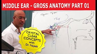
    Excellent method and it is nice to see videos based and focused on concept clearing.
    I really appreciate the effort put in explaining with the help of diagram and not just showing slides.
    Please keep posting more of such videos for nose and throat as well.

  • @ramgopalnagalla4453
    @ramgopalnagalla4453 8 ปีที่แล้ว +14

    thanks sir.that was a beautiful lectures.looking forward to have more lectures.could you please upload inner ear and middle ear anatomy ,physiology and pathology and a a brief about ear

  • @raghunandansirkanungo716
    @raghunandansirkanungo716 9 ปีที่แล้ว +5

    A very Good Lecture delivered by you Thanks a lot

  • @aasiyanadidi8418
    @aasiyanadidi8418 4 ปีที่แล้ว +3

    Great explanation.... Very helpful.. Thank you so much sir..😊🤗

  • @Nikita-wu9ux
    @Nikita-wu9ux 6 ปีที่แล้ว +6

    This video is really helpful....exact wording of dhingra 👏👏

  • @shajudeenps6562
    @shajudeenps6562 5 ปีที่แล้ว +6

    Could you plz add anatomy of eye.your presentatn is simple and useful

  • @abhaygupta1373
    @abhaygupta1373 7 ปีที่แล้ว +1

    sir please taught all the topics of Ear , you videos are so simple does not require more time so please make video on Ear as soon as possible

  • @muhammadshanzaib8802
    @muhammadshanzaib8802 4 ปีที่แล้ว +3

    Thank you so much sir . You are gem 😍😇♥️
    Stay blessed.

  • @KDee755
    @KDee755 6 ปีที่แล้ว +3

    This was a Great Lecture sir

  • @dhruvpatel2529
    @dhruvpatel2529 5 ปีที่แล้ว +2

    Very very nicely and simply explained, please make more videos for anatomy
    Specially on sence organs

  • @ayeshashahzad6238
    @ayeshashahzad6238 2 ปีที่แล้ว +2

    I can't thank you enough sir
    This is the best video ever ❤️❤️❤️❤️

  • @Venugopal-fk3bn
    @Venugopal-fk3bn 4 ปีที่แล้ว +1

    Sir iam from apollo medical clg ur classes r just awesome

  • @amnashahid2920
    @amnashahid2920 ปีที่แล้ว +1

    Very easy and learn able lectures .

  • @meerf
    @meerf 2 ปีที่แล้ว +2

    G bhanu prakash in anatomy and ninjanerd in other subjects
    Are ultimate

  • @medicomuslimahdr
    @medicomuslimahdr 3 ปีที่แล้ว +1

    Absolutely awesome sir . Thanks a lot sir . This lecture ,Really helped me a lot .

  • @tsewangdolma6110
    @tsewangdolma6110 3 ปีที่แล้ว +1

    Very nice explaination sir..thanku very much

  • @sumaiya5065
    @sumaiya5065 6 ปีที่แล้ว +2

    Sir please come to our Allen for teaching.....gr8 work keep it up.

  • @sadiakhan1615
    @sadiakhan1615 7 ปีที่แล้ว +3

    Good class and helpful thanks for uploading ❤

  • @mansurikhalil6774
    @mansurikhalil6774 7 ปีที่แล้ว +1

    very nice lecture sir. it was very informative. we want you to upload more information about middle and inner ear.

  • @patelmeet2008
    @patelmeet2008 4 ปีที่แล้ว +9

    Sir excellent video for understanding, pls upload more video like this in board work of abdomen region

  • @pushpavaliraizada
    @pushpavaliraizada 6 ปีที่แล้ว +3

    Thanks I was eagerly looking for this 💐

  • @ashimrb2511
    @ashimrb2511 6 ปีที่แล้ว +1

    very nice lectures plz upload inner n middle ear

  • @Nihit
    @Nihit 6 ปีที่แล้ว +2

    Very helpful lecture ..please post more on ENT

  • @rajniranjan18
    @rajniranjan18 9 ปีที่แล้ว +10

    i am so thankful sir for this vedio and also i want the same for tme middle ear and inner ear plzz sir make it soon...

  • @Himanshu-pj4wk
    @Himanshu-pj4wk 7 ปีที่แล้ว +2

    thank you sir for this awesome lecture...pls continues making such videos

  • @87saurabchakraborty36
    @87saurabchakraborty36 4 ปีที่แล้ว +1

    Both the videos are awesome

  • @atmo8672
    @atmo8672 4 ปีที่แล้ว +1

    Nice.. said all the relevant points!!

  • @lashmanankv1839
    @lashmanankv1839 5 ปีที่แล้ว +8

    Perfect !!!!!!!!!!!!!!!

  • @danaghazaleh9117
    @danaghazaleh9117 8 ปีที่แล้ว +4

    Thanks a lot. This was well presented

  • @sonijha4109
    @sonijha4109 8 ปีที่แล้ว +2

    awsomely described and told extra point thankyu sir

  • @smartrehan315
    @smartrehan315 5 ปีที่แล้ว +3

    Excellent!! Sir thank you so much..
    I love you sir...

  • @veejetasingh8543
    @veejetasingh8543 4 ปีที่แล้ว +1

    Very awesome video sir can you plz upload video on internal ear

  • @dr.prabhleenkaur8613
    @dr.prabhleenkaur8613 7 ปีที่แล้ว +1

    there is no blood supply and nerve supply . nothing about tympanic membrane . sir please upload a video on this also . i have a exam . nd i understand well from you .

  • @nishachavan4683
    @nishachavan4683 3 หลายเดือนก่อน

    The explanation is so good🥹

  • @mohammadibegum5766
    @mohammadibegum5766 3 ปีที่แล้ว +1

    Plz continue to upload for next topics...

  • @soniababu7193
    @soniababu7193 4 ปีที่แล้ว +3

    Just brilliant. Thank you!!

  • @tarunkasana9749
    @tarunkasana9749 6 ปีที่แล้ว +1

    Very good class sir
    Please do more videos on other topics

  • @Princess-cl6tp
    @Princess-cl6tp 3 ปีที่แล้ว +1

    Bestest video thank you so much

  • @saipriyaj4150
    @saipriyaj4150 9 ปีที่แล้ว +10

    Good class Sir.
    Please do finish by posting videos for middle and inner ear.
    Thank you.

  • @meghnamaumaliya6228
    @meghnamaumaliya6228 6 ปีที่แล้ว +2

    Amazing video sir. Thank you.😊

  • @zoharacuty1430
    @zoharacuty1430 8 ปีที่แล้ว +2

    Thanks a lot sir ..I learned through it

  • @87saurabchakraborty36
    @87saurabchakraborty36 4 ปีที่แล้ว +1

    Sir plz describe the external ear as middle ear

  • @Drmuslimah2019
    @Drmuslimah2019 4 ปีที่แล้ว

    U also explain clinical pts ....that is great

  • @mohammadibegum5766
    @mohammadibegum5766 3 ปีที่แล้ว +1

    Thank you so much sir 🙏🙏🙏🙏awesome explanation

  • @madhumanyam5259
    @madhumanyam5259 3 ปีที่แล้ว +1

    super sir clear voice

  • @priyankajeyaram5235
    @priyankajeyaram5235 2 ปีที่แล้ว +1

    Very good explanation thank u sir

  • @pashamnikitha12
    @pashamnikitha12 ปีที่แล้ว +1

    Thank you sir...🙂🙂

  • @ankitaranisinhatheinnovato6980
    @ankitaranisinhatheinnovato6980 6 ปีที่แล้ว +1

    Superbbbb sir forward the further part

  • @mohdfazal3370
    @mohdfazal3370 4 ปีที่แล้ว

    Sir can u explain , how foreign body gets lodged medial to isthmus of bony part of external acoustic meatus ? bcz it has to pass through isthmus only . why it becomes difficult to remove the foreign body ?

  • @86ujjwalsingh76
    @86ujjwalsingh76 3 ปีที่แล้ว

    Such a nice explanation 🥳
    2nd part??

  • @Hassankhiyal
    @Hassankhiyal 4 ปีที่แล้ว

    Great and helpful work sir ❤️

  • @nagumanbaloch3656
    @nagumanbaloch3656 7 ปีที่แล้ว +1

    tnx sir..please upload more lecture of anatomy

  • @varshamiriyala8699
    @varshamiriyala8699 6 ปีที่แล้ว +2

    pls upload middle ear and inner ear videos sir

  • @priyangaravi2351
    @priyangaravi2351 8 ปีที่แล้ว +2

    awesome lecture sir.. tanx

  • @abduali6680
    @abduali6680 3 ปีที่แล้ว +1

    Great explanation, thanks a lot

  • @thedoctortheguider7464
    @thedoctortheguider7464 ปีที่แล้ว

    Assalam u alaikum sir
    I am unable to find that foramina which you told that is present in anteroinferior of bony canal
    Can you write the name of that foramina please ...

  • @Isumi03
    @Isumi03 ปีที่แล้ว +1

    Perfect!👏❤️

  • @shitalnakiya1513
    @shitalnakiya1513 4 ปีที่แล้ว

    Thank you sir
    That's very fine video

  • @sherondeko1283
    @sherondeko1283 7 ปีที่แล้ว +2

    Many thanx for your knowledge

  • @dancingdoctor4109
    @dancingdoctor4109 3 ปีที่แล้ว

    i was reading scott brown,,, it was so hard to understand.. this helped

  • @lakshmisannala5027
    @lakshmisannala5027 5 ปีที่แล้ว +1

    Nice explanation sir

  • @expressionbyennuu7541
    @expressionbyennuu7541 6 ปีที่แล้ว +1

    Excellent lecture...

  • @dipuno2827
    @dipuno2827 7 ปีที่แล้ว +1

    sir..May you live long.may u can upload more and more videos

  • @angellaturner4584
    @angellaturner4584 7 ปีที่แล้ว +2

    Thanks sir God bless you

  • @minhazislam3273
    @minhazislam3273 3 ปีที่แล้ว +1

    Justtt awesomeeee

  • @abhirockz5508
    @abhirockz5508 7 ปีที่แล้ว

    sir i think you missed tympanic membrane . but the lecture is awesome.

  • @pushpavaliraizada
    @pushpavaliraizada 6 ปีที่แล้ว +1

    Very useful 💚

  • @tanzeelazafar2400
    @tanzeelazafar2400 4 ปีที่แล้ว

    Bestest one👌

  • @bilalmushtaq3810
    @bilalmushtaq3810 2 ปีที่แล้ว +1

    Sir g tusi great o 👌

  • @geojor
    @geojor 9 ปีที่แล้ว +1

    thanks for sharing your expertise...

  • @lavanya5230
    @lavanya5230 4 ปีที่แล้ว +1

    Sir really gd lecture,pls some more videos

  • @priyakanth8472
    @priyakanth8472 5 ปีที่แล้ว +1

    superb sir

  • @kuttykutty...5962
    @kuttykutty...5962 ปีที่แล้ว

    I ask one dout why present in outer ear in our body pls explain

  • @faizbipu1573
    @faizbipu1573 3 ปีที่แล้ว +1

    Thanks a lot

  • @krishnaloveforconsciousnes7284
    @krishnaloveforconsciousnes7284 5 ปีที่แล้ว +1

    Wonderful 🤗🤗🤗🤗😃😃

  • @keepgrinding1198
    @keepgrinding1198 6 ปีที่แล้ว

    Hello Sir, :)
    The auricular conchae is divided further into Cymba conchae and Cavum conchae by the crux of helix.
    Correct me if it’s inaccurate.

    • @doctorbhanuprakash
      @doctorbhanuprakash  6 ปีที่แล้ว

      yess

    • @keepgrinding1198
      @keepgrinding1198 6 ปีที่แล้ว

      Dr.G.Bhanu Prakash Doctor, last year you have visited my college AMEC BCCM in the Philippines with the plan of starting MCI couching classes, I was very much looking forward to it. But there is no more update regarding that. Can you please give an update.

  • @shrutighuntla1158
    @shrutighuntla1158 2 ปีที่แล้ว +1

    thank you sir!

  • @shaikharisha9213
    @shaikharisha9213 4 ปีที่แล้ว

    It was good thanks sir

  • @zameersufiyan327
    @zameersufiyan327 7 ปีที่แล้ว

    sir middle and internal ear anatomy plzz sir ..make it soon

  • @05aishwaryakatwe87
    @05aishwaryakatwe87 4 ปีที่แล้ว +1

    V nice sir upload more videos 😊

  • @ratan2160
    @ratan2160 8 ปีที่แล้ว +3

    thanks sir awasome video

  • @12urvic
    @12urvic 6 ปีที่แล้ว +1

    Sir give me anatomy of middle ear .......and of spinal cord , 3rd ventricle ,4th ventrile...

    • @doctorbhanuprakash
      @doctorbhanuprakash  6 ปีที่แล้ว

      ok sure , to access all lectures visit www.medvizz.com

  • @zainabyounas1141
    @zainabyounas1141 3 ปีที่แล้ว +1

    Best😊

  • @vishnujadhav2345
    @vishnujadhav2345 8 ปีที่แล้ว

    nice lecture sir

  • @vivekchourasiya4688
    @vivekchourasiya4688 5 ปีที่แล้ว

    nice helpful video

  • @satyam7211
    @satyam7211 5 ปีที่แล้ว +1

    Super sir

  • @amolgupta3220
    @amolgupta3220 5 ปีที่แล้ว

    SIR PLZ UPLOAD THE VIDEOS OF MIDDLE AND INNER EAR

  • @aarthimanokaran7197
    @aarthimanokaran7197 5 ปีที่แล้ว +1

    can anyone send the continuation of ths video link

  • @mzafar8177
    @mzafar8177 7 ปีที่แล้ว

    awsum lecture.someone can share the other remaining parts cant find them

  • @Drmuslimah2019
    @Drmuslimah2019 4 ปีที่แล้ว

    Thanks a lot sir

  • @shivanibhardwaj4229
    @shivanibhardwaj4229 6 ปีที่แล้ว

    Sir please give the lacture on internal ear