Wow the amount of knowledge and expertise of an MD Radiologist to read any type of image its astronomical. My respect to you DR. Thank you for showing to us how hard is to read and interpret any illness in the human body.
Please please please do one on CT head!!! I have exams coming up and I'm getting so confused with all the different muscles on CTs !!! Love your channel :)
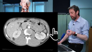
Dr Sam your videos are really good 👍🏼 I think the ureter didn't show up clearly because this CT is an arterial phase rather than an excretion phase where you can visualise them perfectly
Great video as always, Dr Webster. One question: Why are the aorta and vena cava so different in density? Is it just because of the difference in oxygen-content?
Generally CTs are done with IV contrast. This goes directly into the arteries, so they light up more, and during what they call the "arterial phase" of taking a CT the contrast hasn't gotten to the veins yet, so they don't light up as much. The progress of the contrast results in different "phases" that have their different uses for radiologists. This is a helpful video! th-cam.com/video/i9pVTMoRecA/w-d-xo.html


Wow the amount of knowledge and expertise of an MD Radiologist to read any type of image its astronomical. My respect to you DR. Thank you for showing to us how hard is to read and interpret any illness in the human body.
Im always fascinated by imaging, ct, mri, so cool
Please please please do one on CT head!!! I have exams coming up and I'm getting so confused with all the different muscles on CTs !!! Love your channel :)
Dr Sam your videos are really good 👍🏼 I think the ureter didn't show up clearly because this CT is an arterial phase rather than an excretion phase where you can visualise them perfectly
These videos are great. Keep it up! Can you also make videos about cross sectional anatomy (real body slices)?
thank you so much teacher !!!!
Love it! Please do female pelvis! And CT head!!
great video
Can you do one of these of a female pelvis?
do you perform surgeries?
Hello Sam,the captions block the view of your subject. I tried to remove them but it will not turn off
Hm that’s weird, I can turn them off normally
Thanks great help to me make more videos on ct mri images
tq sir
Great video as always, Dr Webster. One question: Why are the aorta and vena cava so different in density? Is it just because of the difference in oxygen-content?
Generally CTs are done with IV contrast. This goes directly into the arteries, so they light up more, and during what they call the "arterial phase" of taking a CT the contrast hasn't gotten to the veins yet, so they don't light up as much. The progress of the contrast results in different "phases" that have their different uses for radiologists. This is a helpful video! th-cam.com/video/i9pVTMoRecA/w-d-xo.html
In this case Aorta is dense because of presence of contrast in it ( arterial phase), contrast hasn't yet reached the inferior vena cava.
That's very good ❤️❤️❤️❤️🌹❤️🌹❤️🌹❤️🌹❤️🌹🌹🌹🌹🌹
🦋
Ureter is normally not seen normally unless there is stone or other pathological conditions
👌👌👌👌👌❤❤❤❤❤❤✔✔✔✔
I don't know why at 23 im learning all the stuff that matters
Hello!
He is Jesus