I am an Engineer and I am 80 years of age. I have viewed number of videos on ECG and also referred to a few text books. After following for a while I get lost and stop further reading or postpone watching. This video is so clear that I could follow till the end without fatigue. Looks like this is created by a technical person. it is excellent.
Never saw anything like this whole career of 30 years. Kudos to you. Explanations like this in medical school could make the life of students so simple. Please keep up the fantastic work. 🙏🙏
Wow! I am a seccond year medical student and I could not understand anything from my professor(because he just read the slides) and I felt that I am idiot but thanks to you I understand everything. Thanks a lot!!
This is the BEST video on the basics of EKG I have ever seen. None of the sources I saw would explain why the leads show their particular wave pattern in detail. I didn't really understand it until you explained the direction of depolarization in each segment of the myocardium and how the electrodes "see" these changes dependent on the lead's angle. Thank you thank you thank you for explaining this so beautifully. Teaching really is an art form and you are a master of the art!
I have been a RN for about 1.5 years and wanted a deeper understanding of EKG readings. This was AMAZING. So clearly explained and so easy to follow visually. This has helped me so much, and I look forward to using this in my clinical practice. THANK YOU!!!!!
This was amazing, I like how you built up our knowledge to understand everything. From how electrical potential is recorded to direction of changes of potential to the entire ecg. Thank you for making this.
That makes so much sense. I memorized many different interpretations of EKG abnormalities, but I always felt like I didn't really grasp the basics, even after watching tons of videos and reading a lot. You explained it so clearly, literally made me cry for joy. Thank you so much!
This demo is Not only for RPN,DR's or students.Everyone who is interested in GK must watch and digest this video.This subject has never been demostrated like this in any Lecture or vedios.Thanks to the originator.
I'm not a healthcare professional but wanted to understand the rationale behind the ECG science.... Never seen such nicely and detailed explanation of the 12 lead ECG theory. Love it!
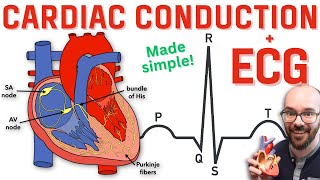
Good description. I like to add one thing about the AV node. The isoelectronic line represening the AV node conduction represents one more thing than simply the lack of potential. It represents the delay in conduction caused by the slow calcium channel instead of the fast sodium channels in the other parts of the heart. This delay is called 'the nodal delay' that is needed to give the ventricles a time to dilate while the atria are contracting. Otherwise, the atria and ventricles would contract simultaneously and the blood will not go to anywhere from anywhere. Thank you.
Amazing video! great job! One of the best ECG videos i EVER watch. If you continue to break down medical topics as basic and easy to understand and make this your “trademark voice”, i bet you will gain so much more subscribers. Your channel is a hidden gem 💎 I Subscribed for sure. And I LOVE the animation!!! 👍
This will be an excellent learning material for my students to have a good fundamental on the basic physiologic principles of ECG. I'm certain that they will benefit from it.
Great thank to you. That makes me understand better on ECG than reading text books. Please continue with other medicals videos for our medical students. Thank you again❤❤❤❤❤❤❤
Thanks sir, for this kind of hard work and sharing this beautifully, simplified and oriented educational ECG lecture which I understood after so many months. Really it's very easy for newly medico learners and of course revision for many.
Amazing I have seen many videos on ECG basics but some confusions always remained uncleared like the direction of propagation of repolarization in ventricular compartment. Physiology of Q wave formation. Concept behind lead placements and most important and basic one physiology of measuring electrical activity in relation to these conducting cells and propagation of impulse. I mean every confusion is cleared in a single video which was not cleared after watching every top of the list videos on ECG on TH-cam which was like watching period of about 5 to 6 hours. Best of the best. Please keep making these kind of videos and don't worry about length of video because conceptual study students don't see length of video when they are watching videos like this. Stay blessed and Keep making more videos ❤❤❤❤❤❤
ABSOLUTELY FANTASTIC VIDEO, EVERY SINGLE SECONDD MOMENT OF THIS VIDEO WAS PRECIOUS, TREASURE TROVE OF INFORMATION AND UNDERSTANDING, INDEEEEDDDDD THE VIDEO WAS JUST LIKE THE WAY YOU DESCRIBED IN THE INTRODUCTORY PART LOVE LOVE LOVEEE SUPERRRRR LOVEDDDDDD ITTTTTT WITH ALL MY ECG WITH ALLL MYYYYY HEART❤❤❤❤❤❤❤❤❤❤❤❤❤❤❤❤❤❤
Super!! I did my notes , graphs and sketches time ago to understand it as it was explained here , but i lost them like twice and was lazy to repeat it hahha so you save me some time to refresh my memory from time to time😁
I’ve seen many presentations on this subject but this one is very well done and easy to follow. The shared “electrode” in the middle of the heart I’ve always known as a vector derived “central terminal”
AvR lead looks from the right shoulder and it sees the positive depolarising wave moving left side.Means DP is moving away,hence QRS is negative in aVR.Even in V1 the R wave is very small compared to V6.If aVR is positive it either means lead placement has been reversed or it's a case of dextrocardia.
This is a fantastic video and one of the few that show why we get a negative Q-wave in the QRS complex, I love the animations as well. I would like to add something however, and that is in you animation demonstrating the cardiac cycle you are showing negative charges spreading from the atria to the ventricles implying that the resting membrane potential of the cells is inherently positive which is inaccurate because its the positive charges that spread downward while the cells get depolarises(from neg to pos) and the charges moving towards the positive electrode producing an upward deflection...


I am a practicing Dr and I have never seen such a nice video ever on ECG , congratulations 🎉🎉🎉
Hi
Where are you from
I am from U.P state of india
Awesome
I am a cardiologist resident and I can say this is far one of the best videos on youtube.
I am an Engineer and I am 80 years of age. I have viewed number of videos on ECG and also referred to a few text books. After following for a while I get lost and stop further reading or postpone watching. This video is so clear that I could follow till the end without fatigue. Looks like this is created by a technical person. it is excellent.
The same goes with me....but I am 46 only....
Never saw anything like this whole career of 30 years. Kudos to you. Explanations like this in medical school could make the life of students so simple. Please keep up the fantastic work. 🙏🙏
Wow! I am a seccond year medical student and I could not understand anything from my professor(because he just read the slides) and I felt that I am idiot but thanks to you I understand everything. Thanks a lot!!
Same 😅
Same for me and just watching this I understand way better than Dr. S just reading the slides🤦🏽♀️
Different country,Same professor.
Nowadays, most of the professors are slide readers, not explain properly. This is the most best video ever I have found.😅
You're the best man.... Nobody will be able to explain like that.... Thank you so much. 😮
This is the BEST video on the basics of EKG I have ever seen. None of the sources I saw would explain why the leads show their particular wave pattern in detail. I didn't really understand it until you explained the direction of depolarization in each segment of the myocardium and how the electrodes "see" these changes dependent on the lead's angle. Thank you thank you thank you for explaining this so beautifully. Teaching really is an art form and you are a master of the art!
Has to be one of the best explanation of ecg. Better than any text book I've read
I am registered nurse. This is a amazing video to get to know about basics in ECG.
I have been a RN for about 1.5 years and wanted a deeper understanding of EKG readings. This was AMAZING. So clearly explained and so easy to follow visually. This has helped me so much, and I look forward to using this in my clinical practice. THANK YOU!!!!!
The best video about ecg basics 👌
This was amazing, I like how you built up our knowledge to understand everything. From how electrical potential is recorded to direction of changes of potential to the entire ecg. Thank you for making this.
That makes so much sense. I memorized many different interpretations of EKG abnormalities, but I always felt like I didn't really grasp the basics, even after watching tons of videos and reading a lot. You explained it so clearly, literally made me cry for joy. Thank you so much!
Glad to hear that
I mean "you finally understood" part😅
This demo is Not only for RPN,DR's or students.Everyone who is interested in GK must watch and digest this video.This subject has never been demostrated like this in any Lecture or vedios.Thanks to the originator.
I don´t know how long I searched for a video with such quality of information. Thank you so munch!!
You deserve all the accolades other viewers have already showered upon you. Thank you so much for such an amazing and wonderful educational video.
I'm not a healthcare professional but wanted to understand the rationale behind the ECG science.... Never seen such nicely and detailed explanation of the 12 lead ECG theory. Love it!
OMG all doubts cleared in one go
The visuals are awesome! Thank you for such an awesome video! ❤
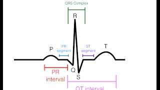
Good description. I like to add one thing about the AV node. The isoelectronic line represening the AV node conduction represents one more thing than simply the lack of potential. It represents the delay in conduction caused by the slow calcium channel instead of the fast sodium channels in the other parts of the heart. This delay is called 'the nodal delay' that is needed to give the ventricles a time to dilate while the atria are contracting. Otherwise, the atria and ventricles would contract simultaneously and the blood will not go to anywhere from anywhere. Thank you.
Thank you sir ❤
Thank you so much ❤
I am a 1st medical student. I was in big confusion with this ECG , and now I have a clear 💡 idea.
I am supposed to take a test tomorrow and this helped me and my colleagues. Thanks a lot!!!❤
Totally flawless ❤ to the point and crystal clear.
Thanks for making this concept very easy and interesting 💫💎.
Huge thanks, love all your explanations
This one is huge❤thx a lot for this one.
Besttttttt video....short and fully packed...nice job sir
Comparatively the best Ecg explanation in TH-cam !
Thanks!
Thank you for the super thanks. It really helps.
Part two . About irregularities
Also video on CT scan , ultrasound
Yes plz!!!
Thanks so much from🎉🎉🎉 an ER‐nurse from germany
Excellent. Never found such an thorough explanation.
I'm a first-year medical student. This channel's gonna help me a lot 😆
Welcome...
🔥🔥best explanation I've ever seen for ECG basics
Really really really it is the best explanation you could find on youtube❤
It’s been a great pleasure to listen to this amazing man!
Its really wonderful...i am an engineer and inderstood the ECG concept today only ..thanks for such an excellent video
This is very well presented how electrical knowledge used in cardiovascular detection. Thank you.
_So_ well done. Biology made clear to an electrical engineer!
Such a great video, the only one that made me understand where the waves in the ECG come from. Thank you so much!! :D
Amazing video! great job! One of the best ECG videos i EVER watch. If you continue to break down medical topics as basic and easy to understand and make this your “trademark voice”, i bet you will gain so much more subscribers. Your channel is a hidden gem 💎 I Subscribed for sure. And I LOVE the animation!!! 👍
This will be an excellent learning material for my students to have a good fundamental on the basic physiologic principles of ECG. I'm certain that they will benefit from it.
This explanation is awesome! Excellent!
Thank you sir ❤️❤️ for great explanation.
I am a pharmacist and this really helped me cause l have a physiology exam
This is ganna be viral.
The best explanation of EKG concept, as an engineer.
Great thank to you. That makes me understand better on ECG than reading text books. Please continue with other medicals videos for our medical students. Thank you again❤❤❤❤❤❤❤
Thanks sir, for this kind of hard work and sharing this beautifully, simplified and oriented educational ECG lecture which I understood after so many months. Really it's very easy for newly medico learners and of course revision for many.
Greetings from Nairobi and many thanks for your educative video. I have learned a lot.
This is the best Video so far. Thank you Dr.
I have never seen such a great explanation! Amazing! Great job.
4:45 yeah it's really very good i wanna cryyyyyyy soooooo many prayers for youuuuuuu❤❤❤❤❤❤❤❤❤❤❤❤❤❤❤❤❤❤❤
Very easy explanation to understand what is ECG .duration between one circle is 0.83 second.it is normal heart beat.thank you
This was very informative. Thank you!
Amazing I have seen many videos on ECG basics but some confusions always remained uncleared like the direction of propagation of repolarization in ventricular compartment. Physiology of Q wave formation. Concept behind lead placements and most important and basic one physiology of measuring electrical activity in relation to these conducting cells and propagation of impulse. I mean every confusion is cleared in a single video which was not cleared after watching every top of the list videos on ECG on TH-cam which was like watching period of about 5 to 6 hours. Best of the best. Please keep making these kind of videos and don't worry about length of video because conceptual study students don't see length of video when they are watching videos like this. Stay blessed and Keep making more videos ❤❤❤❤❤❤
First time i have deeply understood the concept of ecg,Thanks alot
Incredible explanation.
what an explanation ....great sir
This is the best ecg video I have ever seen ❤️❤️❤️❤️❤️❤️
Good explanations . Thank you sir.
Such an amazing video...thanks for making my concepts clear in such less time.
Very nice explanation. Highly appreciated👍👍
Thank you for that amazing explanation👍🏻👍🏻👍🏻
ABSOLUTELY FANTASTIC VIDEO, EVERY SINGLE SECONDD MOMENT OF THIS VIDEO WAS PRECIOUS, TREASURE TROVE OF INFORMATION AND UNDERSTANDING, INDEEEEDDDDD THE VIDEO WAS JUST LIKE THE WAY YOU DESCRIBED IN THE INTRODUCTORY PART LOVE LOVE LOVEEE SUPERRRRR LOVEDDDDDD ITTTTTT WITH ALL MY ECG WITH ALLL MYYYYY HEART❤❤❤❤❤❤❤❤❤❤❤❤❤❤❤❤❤❤
Wow, thank you!
Super!! I did my notes , graphs and sketches time ago to understand it as it was explained here , but i lost them like twice and was lazy to repeat it hahha so you save me some time to refresh my memory from time to time😁
Marvelous explanation of such a difficult topic.
Thanks a lot Sir.
With Love, Lot of Appreciations and Best Regards from Pakistan
Good job 👏👏 very clear description
Brilliant video. Absolutely brilliant.
Thankyou so much for this amazing video... Understood the concepts in one go..❤️
Great great job with so much simplified manner of a very tough topic,Thanks.
The best video on ecg in history
Thank you so much for the appreciation
Excellent description understood well all about ECG many many thanks looking forward to seeing more videos
This was explained so well! I am so glad I came across this video. Thank you so much for creating this!
Noone would match the level of this video.....huge thanksssss
Thank you doctor you helped alot of lectures
super- clear explanation--- great job.
I’ve seen many presentations on this subject but this one is very well done and easy to follow. The shared “electrode” in the middle of the heart I’ve always known as a vector derived “central terminal”
It is actually helpful. Thank you so much!
Wonderful explaination👍
Understanding first time the ecg formation
👍👍🙏🙏
Very well explained! Thank you so much
Excellent lecture......
Thanks so much for sharing your knowledge with us. I appreciate you! ❤
really informative..
Thanks. I can tell you put a lot of heart into this lesson. Most excellent. 🙄
Bro 🔥🔥🔥🎉🎉🎉❤️🔥❤️🔥❤️🔥
This is absolute Cinema 🙌🙌
Wow! Im slowly getting it, 😅 Thank you so much for this content. I will be following you from now on❤
This video is so useful thank you so much
The best video that I have ever see n . Explaination was clear and amzing. Thank you bro
Thanks, it is very clear explanation On ECG .I was seen many videos concerning ECG but this is very interesting one.
Simply excellent presentation.
it was immensely useful... thank you for making it interesting.your efforts really made a change in the way i learn
Very well explained, thank you
thank you for simplified explanation😄
Please share.
god bless you sir this was amazing
Please do common cardiovascular disease on ECG in this same way
I'm searching for someone to explain the pathology regarding its electrical changes
setelah sekian purnama akhirnya paham, makasih🥰
AvR lead looks from the right shoulder and it sees the positive depolarising wave moving left side.Means DP is moving away,hence QRS is negative in aVR.Even in V1 the R wave is very small compared to V6.If aVR is positive it either means lead placement has been reversed or it's a case of dextrocardia.
This is a fantastic video and one of the few that show why we get a negative Q-wave in the QRS complex, I love the animations as well. I would like to add something however, and that is in you animation demonstrating the cardiac cycle you are showing negative charges spreading from the atria to the ventricles implying that the resting membrane potential of the cells is inherently positive which is inaccurate because its the positive charges that spread downward while the cells get depolarises(from neg to pos) and the charges moving towards the positive electrode producing an upward deflection...
It was one of the best videos ab😢ECG out