Evertime you start any embryology lecture, you often revise from the beginning... I find it really useful sir... It helps me to recall what I have learnt before...Thank you so much Sir...❤ from 🇮🇳
@@HayaEsmaiel-uu8uo مرحباً! يمكنك مشاهدة الفيديوهات بالترتيب الصحيح من خلال قائمة التشغيل المنظمة هنا: th-cam.com/play/PLnkPtP3elLY6SLpfiQNWHB0FIGeTDxLbH.html القائمة مرتبة بشكل تسلسلي منطقي يتبع تطور التجاويف الجنينية خطوة بخطوة. ننصحك بمشاهدة الفيديوهات بالترتيب الموجود في القائمة للحصول على أفضل فهم للموضوع. استمتع بالتعلم! 🎓
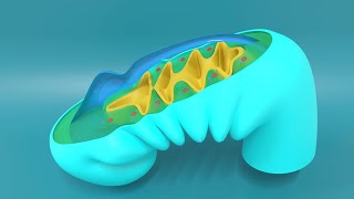
Intrembryonic mesoderms are continuous with the splanchnic of extraembryonic mesoderm. Since the somatic extraembryonic mesoderm is only continuous to the splanchnic extraembryonic mesoderm by connecting stock. Please check this out
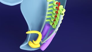
Actually this is not the perfect model. At buccopharyngeal membrane and cloacal membrane, there is little to no mesoderm. Thank you so much for pointing this out. I should have fixed it in the model. But since this was not the main topic, it was overlooked. I am sorry.
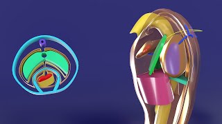
Sir at 8:12 , the cranial part of embryo ( near oropharyngeal memb ) have paraxial mesoderm , intermediate mesoderm and lateral plate mesoderm at midline of cranio-caudal axis . So ,this means the intra embryonic mesoderm will be lateral upto presence of notochord . Then , it move towards midline covering notochord cranially . Am I correct sir
Thanks for watching and leaving a comment! Please let me know for what purpose do you need the .blend file? Feel free to contact me directly via any one of the following channels: 1. Live chat at www.medicovisual.com 2. WhatsApp text at +923037241030 3. Email at draizaz@medicovisual.com 4. Contact us form at www.medicovisual.com Best regards, Dr. Aizaz
How's it possible that in a longitudinal section all the three portion of mesoderm i. e., The paraxial mesoderm, the intermediate mesoderm and lateral plate mesoderm are visualized???
I am sorry, I didn't quite understand your question. Can you please elaborate how and why all three portions of mesoderm cannot be visualized. Also please include the timestamps of the video and if necessary, references from a book.
@@MedicoVisual I'm saying in midline is notochord, slightly lateral to it's paraxial mesoderm, then intermediate mesoderm and lateral most is lateral mesoderm,,, so if we take longitudinal section of Embryo will we be able to see all the three division of mesoderm in longitudinal section? It's not possible... But, it's possible to see 8n transverse section
@@MedicoVisual Sir, I might be wrong...But the reason why I'm saying we can't visualize all the three mesodermal portions in longitudinal section is that they're not all present in some longitudinal plane... Paraxial mesoderm being medial most, slightly lateral to it's intermediate mesoderm, while lateral most is Lateral mesoderm,,,, so if I think a longitudinal section I can't see all the three.... Please do correct me if I'm wrong!
Not at all intuitive.. Messed up everything Ur just complicating that topic Dont zoom past, no clear visual of coleomic cavities during folding, no ecto, endoderm and extra embryonic mesoderm during folding......
Dear @MuthuThala-ih2wu, Thank you for your feedback on my lecture. I apologize if the explanations and visuals were not clear or intuitive enough. Your input helps me identify areas for improvement. I will review the sections you mentioned and work on providing more detailed and understandable explanations in future lectures, particularly regarding the coelomic cavities, embryonic folding, and the roles of the germ layers. If you have any specific suggestions or questions, please feel free to share them. I appreciate your engagement and am committed to supporting your learning journey. Best regards, Dr. Aizaz
@@MedicoVisual include extra embryonic mesoderm, ectoderm, endoderm during embryonic folding. After 20 mins video got too complicated bcoz of u not explaining about it properly. All ur videos are Intuitive, except these embryonic foldings...
@@MuthuThala-ih2wu Thank you for your comments. I will try to create separate lectures specifically on the Embryonic folding that will include all the structures in the animation.


Enroll in complete course for FREE to access practice MCQs
www.medicovisual.com/courses/premium-3d-body-cavities-diaphragm-embryology
how can i get MCQS
@@endex-s6yPlease Enroll into the free course. They are inside the members area.
Evertime you start any embryology lecture, you often revise from the beginning... I find it really useful sir... It helps me to recall what I have learnt before...Thank you so much Sir...❤ from 🇮🇳
these videos saved my life!!! thank you doctor. mashallah
This is the best way to teach embryology
These videos had clear all my doubts😊😊
Glad to hear that
Hello. I'm a medical student from Russia, and I love your videos. Thanks you so mutch, you're a wonderful lector
о, я тож из России
@@agersolt818 я из Омска, лечфак, 3 курс
@@vicktorshushackov3074 Я из Москвы, леч, 4 курс
@@vicktorshushackov3074пытаешься тоже вникнуть в эмбриологию?
@@agersolt818 ага, для себя. Мы уже сдали её, но на парах не очень понял
Best ever animation and explanation..
Most effective way of teaching
Thanks a ton
Thank you ❤
Love from Iran ❤
You are a LEGEND!!!!!!
Thank you so much.
كيف اعرف الترتيب الافضل للفيديوهات ؟الشرح فوق الخيال
@@HayaEsmaiel-uu8uo مرحباً! يمكنك مشاهدة الفيديوهات بالترتيب الصحيح من خلال قائمة التشغيل المنظمة هنا:
th-cam.com/play/PLnkPtP3elLY6SLpfiQNWHB0FIGeTDxLbH.html
القائمة مرتبة بشكل تسلسلي منطقي يتبع تطور التجاويف الجنينية خطوة بخطوة. ننصحك بمشاهدة الفيديوهات بالترتيب الموجود في القائمة للحصول على أفضل فهم للموضوع.
استمتع بالتعلم! 🎓
Intrembryonic mesoderms are continuous with the splanchnic of extraembryonic mesoderm.
Since the somatic extraembryonic mesoderm is only continuous to the splanchnic extraembryonic mesoderm by connecting stock. Please check this out
Thank you so much for pointing it out.
Can you please share a relevant reference?
So intra embryonic splenchnic mesoderm as well as intra embryonic somatic mesoderm both continues with extra embryonic splenchnic mesoderm right?
@@zebra4387yes, but through the connecting stalk
@Medicovisual sir this means at 7.37 min mesoderm is invaginating buccopharyngeal membrane also
Actually this is not the perfect model. At buccopharyngeal membrane and cloacal membrane, there is little to no mesoderm. Thank you so much for pointing this out. I should have fixed it in the model. But since this was not the main topic, it was overlooked. I am sorry.
Excellent lecture Dr Aizaz 👌
Nice Presentation
Hello sir in which lecture u discussed about septum transversum ?
In this lecture from 26:26 onward and in GIT Embryology course with development of liver.
Nice illustrations
excellent video
Thank you sir ❤
Sir at 8:12 , the cranial part of embryo ( near oropharyngeal memb ) have paraxial mesoderm , intermediate mesoderm and lateral plate mesoderm at midline of cranio-caudal axis . So ,this means the intra embryonic mesoderm will be lateral upto presence of notochord . Then , it move towards midline covering notochord cranially . Am I correct sir
Yes that correct. There are right and left limbs of mesoderm that converge in the cranial midline to form unfused cranial midline part.
Genius even better than the texts
Thank you, Dr. Aizaz!
Is it possible to share the blender file, this is too amazing to miss out on
Thanks for watching and leaving a comment!
Please let me know for what purpose do you need the .blend file?
Feel free to contact me directly via any one of the following channels:
1. Live chat at www.medicovisual.com
2. WhatsApp text at +923037241030
3. Email at draizaz@medicovisual.com
4. Contact us form at www.medicovisual.com
Best regards,
Dr. Aizaz
Did you do a graphics design degree before the medical degree dr? 😮
No. I do not have a graphics design degree. I self-learned blender from TH-cam and Udemy.
Sir how can we get your 3d sofmodels🥺
I am sorry. These 3D models aren't available, as of now.
What is the name of the app that you used in the video ?????
@@lianebans7885 Blender 3D
Thanks for this lesson
Always welcome
Sir is the diaphragm inside the coelom?
@@anjalaansari6082 Yes
What is the name of the application that you are using ??
Blender (www.blender.org)
How's it possible that in a longitudinal section all the three portion of mesoderm i. e., The paraxial mesoderm, the intermediate mesoderm and lateral plate mesoderm are visualized???
I am sorry, I didn't quite understand your question. Can you please elaborate how and why all three portions of mesoderm cannot be visualized. Also please include the timestamps of the video and if necessary, references from a book.
@@MedicoVisual I'm saying in midline is notochord, slightly lateral to it's paraxial mesoderm, then intermediate mesoderm and lateral most is lateral mesoderm,,, so if we take longitudinal section of Embryo will we be able to see all the three division of mesoderm in longitudinal section? It's not possible... But, it's possible to see 8n transverse section
During 8-9 minutes.... During craniocaudal folding
@@akbashirahmad okay I got you. But why do you think it's not possible to visualize different parts of mesoderm in this section?
@@MedicoVisual Sir, I might be wrong...But the reason why I'm saying we can't visualize all the three mesodermal portions in longitudinal section is that they're not all present in some longitudinal plane... Paraxial mesoderm being medial most, slightly lateral to it's intermediate mesoderm, while lateral most is Lateral mesoderm,,,, so if I think a longitudinal section I can't see all the three....
Please do correct me if I'm wrong!
Thx
Thank you very much 😊
I also wanna become a doctor.
Most welcome 😊
What is name of this app ?😮
It's a software called "Blender 3D"
This program name please?❤
Blender 3D
10.45: transverse section or coronal section?
Transverse section
❤❤❤
What that app you have used?!
It's a software called "Blender"
❤
🤩
Not at all intuitive..
Messed up everything
Ur just complicating that topic
Dont zoom past, no clear visual of coleomic cavities during folding, no ecto, endoderm and extra embryonic mesoderm during folding......
Dear @MuthuThala-ih2wu,
Thank you for your feedback on my lecture. I apologize if the explanations and visuals were not clear or intuitive enough. Your input helps me identify areas for improvement.
I will review the sections you mentioned and work on providing more detailed and understandable explanations in future lectures, particularly regarding the coelomic cavities, embryonic folding, and the roles of the germ layers.
If you have any specific suggestions or questions, please feel free to share them. I appreciate your engagement and am committed to supporting your learning journey.
Best regards,
Dr. Aizaz
@@MedicoVisual include extra embryonic mesoderm, ectoderm, endoderm during embryonic folding.
After 20 mins video got too complicated bcoz of u not explaining about it properly.
All ur videos are Intuitive, except these embryonic foldings...
@@MuthuThala-ih2wu Thank you for your comments.
I will try to create separate lectures specifically on the Embryonic folding that will include all the structures in the animation.
What is the name of the app that you used in the video ?????
@@lianebans7885 Blender 3D