The Infratemporal Fossa - Boundaries & Contents | Anatomy Tutorial

ฝัง
- เผยแพร่เมื่อ 2 ต.ค. 2024
- #infratemporal #maxillary #mandibular
Donation Link: paypal.me/stud...
/ anatomy.knowledge
The mandibular nerve exits the skull through foramen ovale. Just under the foramen ovale its gives off a recurrent meningeal branch called the nervus spinosum which enters the skull thorugh foramen spinosum. The otic gg. Is also attached to the mandibular nerve at this level.
Soon the mandibular nerve splits into an anterior and a posterior division.
From the anterior division are arising branches like the : deep temporal nerves, masseteric nerve and buccal nerve.
The posterior division of the mandibular nerves splits into three branches:
The inferior alveolar nerve which goes inferiorly to enter the mandibular foramen;
The lingual nerve which goes anteroinferiroly;
And the auriculotemporal nerve which arises by two roots from the posterior division of the mandibular nerve.
The chorda tympani nerve, a branch of the facial nerve, appears in the infratemporal fossa and unites with the lingual nerve.
The maxillary artery is one of the two terminal branches of the external carotid artery. The other terminal branch of external carotid is the superficial temporal artery.
The maxillary artery arises at a straight angle from the external carotid, passes medial to the neck of the mandibular condyle and goes anteriorly to leave the infratemporal fossa by way of pterygomaxillary fissure. It gives numerous branches like the Middle meningeal artery which goes upwards, passes inbetween the two roots of auriculotemporal nerve and enters the skull by way of foramen spinosum. An accessory meningeal artery may arise to enter the skull by oway of foramen ovale.
The inferior alveolar artery follow the course of inferior alveolar nerve;
Other branches from the maxillary artery are:
The masseteric artery, buccal artery and the anterior and posterior deep temporal arteries.
The Pterygoid venous plexus accompanies the maxillary artery and towards the neck of the mandibular condyle gives rise to the maxillary vein. The maxillary vein unites with the superficial temporal vein to form the retromandibular vein.
The muscular structures present in the infratemporal fossa are the :Lateral & medial pterygoid muscles , & tendon of temporalis;


![เปิดบ้าน พี่ตูน Bodyslam หลังใหม่คนแรก ที่ภูเก็ต l [Nickynachat]](http://i.ytimg.com/vi/flFwcmyDQSk/mqdefault.jpg)


you are the best teacher sir!!! No youtube medical channel can match your way of teaching and precision!!
Sooper class....Tnk uh so much sir...The best way of teaching I have ever heard...keep going sir...
OMG, you can't even imagine how this video changes my life!! thank you
After so many lectures of my friends,professor, i clearly understood this now😅
Wonderful lecture .. thank you sir❤
Wow. You're ambidextrous. Thank you so much for this master piece.
Can't thank you enough for this! Much appreciated efforts
Brilliantly explained, I can't thank you enough for this ❤️❤️
Thank you so much! Very helpful!
Amazing explanation. Thank you very much
🤩 brilliant
Thank you so much for this.. U r truly amazing
Brilliant, thank you! Please continoue uploading as your videos are helping lots of students like me.
I will! Thank you!
Masterpiece!
Very useful video
Sir, please upload more head and neck anatomy videos
Thank you for your hard work. The description box is awesome as well, thank you :)
He writes in both hands ✍ 😲
Excellent!
You're the best, sir! 👏👏🤗
Chorda tympani is a branch of facial
Thank you for the video! The description box is not updated :)
Amazing video!!! Your video literally forced me to click the subscribe button
simply beautiful ...i couldnt resist subscribing to your channel....kudos
Thanks alot
You got another subscriber brother. This is brilliant ❤️
Thank you very much!
Wow
Thank you verrryyy muchhh mate
wowwww .... explained everything with such an ease
Thank you!
This video covered whatever was written in the Infratemporal fossa of the Clinically Oriented Anatomy textbook.
The description from ur diagram is brilliant 🌟🌟
wow!!! This was the best explanation ever!!
Wow
Very Nice thank you very much 😊
A new subscriber here, thanks alot sir, it was very informative:)
sir you are teaching very nicely at any moment i didnt feel bore thank so much sir for giving this much of knowledge for us thank you so much sir
❤️❤️💙💙
this is the best youtube channel for revising anatomy
thank you so much sir, with the help of your videos its not only easy to practice the diagrams but also for quick revision. your video
helped me over come the fear of the topic.
Sir tell more about yourself
Well explained 🌟🌟🌟
Wow how comes i encountered this page late🥺
رهيب الشرح❤❤❤
Thank you
Wow! Thank you so muchh ❤️❤️
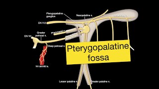
As usual awesome video.. bro, please post pterygopalatine fossa anatomy if possible..
Thank you! It is already posted th-cam.com/video/QacrWgdmzdY/w-d-xo.html
Nive video
This is amazing i can't describe how i fell now. You explained it very cleanly and beautifully.Thank you for your effort:)
Thank you for making these complex topics easier!!!
glad to find this before preproffssss🥹🥹❤️