Liver Ultrasound Probe Positioning | Transducer Placement For Liver Scanning | Abdominal USG

ฝัง
- เผยแพร่เมื่อ 28 พ.ค. 2024
- Liver Ultrasound Probe Positioning | Transducer Placement For Liver Scanning | Abdominal USG
*Timestamps:
Intro - 0:00
Left Lobe Of Liver - 0:08
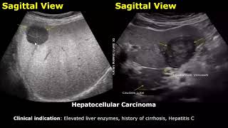
Caudate Lobe - 2:29
Porta Hepatis & Bile Duct - 3:07
Right Lobe Medial To Kidney - 4:02
Right Lobe And Kidney - 4:29
Right Lobe And Hepatic Veins - 5:08
Liver At Porta Hepatis Level - 6:22
Liver And Kidney - 6:59


![[UNCUT] รัฐสั่งหยุดปล่อยคลื่น! แต่หยุดพลังปะทุ “น้องหญิง” ไม่ได้ I คนดังนั่งเคลียร์](http://i.ytimg.com/vi/RmQh47eIiV0/mqdefault.jpg)


How To Measure Liver On Ultrasound: th-cam.com/video/A3akbgBKmcw/w-d-xo.html
Liver Ultrasound Normal Vs Abnormal Image Appearances: th-cam.com/video/-0WrCtDe6IQ/w-d-xo.html
What I love the most about this video is the probe placement. It is extremely helpful. I hope we would get more videos like this, especially for an obstetrics ultrasound scan. Thank you
Thank you so much for watching! Here is the video on obstetric ultrasound probe positioning. More videos on it will be uploaded in the future. th-cam.com/video/bRPD-51UG7Y/w-d-xo.html
Sir please show diagramatic views as well with the probe positioning. I'm not able to correlate with the ultrasound images. Please 🙏 sir
I agree with you. With multiple ultrasound videos hardly anybody explains how to position and angle probe to obtain them. Dr. Sam is thorough and detailed, and explains everything very clearly.
It's always a pleasure to learn through Indian doctors videos. You are by far the best. Big Thanks from Morocco for sharing
Thank you very much for watching! Greetings!
dr sam what a beautiful lecture,pls continue.
Thank you so much for watching!
Extremely helpful video tysm!
Most Welcome!
Very helpful!!! Thank you!
Most Welcome!
I love your videos thanks!!
Thank you very much for watching!
Thank you sincerely for all your videos.
Most Welcome and thanks for watching!
Thank you,doctor Sam.
Most Welcome!
Very benificial. That's what I was searching
Thanks for watching! Glad to hear it!
Well explained. Thank you Dr
Most Welcome!
Thank you. Very educating
Most Welcome!
Excellent thanks sir
Most Welcome!
Amazing video... Congratulations for your spiritual thought..
Thank you very much for watching!
Thank you! This is very helpfull
Most Welcome!
Thank you very much Dr
Most Welcome!
How can we change the color of the screen in this machin ?
Congratulation! Very good presentation. Next time I am waiting for a manner of measurement of all the figures of the liver (e.g. left hepatic lobe, pv, cbp etc). Cristi/Romania. All the best!
Thank you very much for watching! Yes, liver measurements video has been uploaded. Here is the link: th-cam.com/video/A3akbgBKmcw/w-d-xo.html
Thanks a lot!
@@cristiariesan8320 Most Welcome!
Thank u so much
Dear teacher 🥰🙏
Most Welcome!
Can ulcers of colon can be seen in sonography or ct scan?
Thank you.
Most Welcome!
Thank you so much sir 😊🙏
Most Welcome!
Thank you
Most Welcome!
sr liver se related bimari bhj explain kiya kro please
You can watch it here: th-cam.com/video/-0WrCtDe6IQ/w-d-xo.html
I have to go for a ultrasound on my l8ver Friday hopefully its not bad tho
Thanks alot sir
Most Welcome!
Thank you Sir
Most Welcome!
Why is there no mention of Intercoastal view?
Nice presentation sir
Thank you very much for watching!
Very helpful
Thanks for watching!
Why is there no mention of Intercoastal section ??? Or is it not necessary in all patients????
Nice
Thanks for watching!
Thanks sir
Most Welcome!
Tnx a lot
Most Welcome!
Wonderful sir.
Dear sir, facing difficulties while scanning kidney, particularly while confusing fat in renal sinus with stone. Sometimes in presence of flank pain, one errs to report fat with stone.
Kindly educate more
Thanks for watching! Fat will not cause posterior shadowing whereas most stones will cause posterior shadowing.
Can be video the location porb all the organ please
Yes, they will be uploaded in the future.
Also obestric and gynecology
Thanks for watching! Yes, sure!
Is there a dedicated video for imaging the IVC?
It will be uploaded in the future
@@DrSamsImagingLibrary thank you. I eagerly await. It helps me with my clinical practice as an ultrasound student.
Dear teacher, how to measure complete liver length???
The craniocaudal length is mainly used. You can watch this video for liver measurements. Thanks. th-cam.com/video/A3akbgBKmcw/w-d-xo.html
Doc, is it possible to interpret ultrasound wrongly in liver tumor?
Yes, such misdiagnosis can happen in an ultrasound scan
Plz do Obs & gyne sir
Thanks for watching! Yes, sure!
Sir cirrhosis can detect usg?
Yes it can be detected
Pancreas k liye bhi Sir.
Thanks for watching! Yes, sure!
Plz real time scan main images and measurements lena seekha den sary organs ki..plzz
Thanks for watching, I will try.
Hii sir g Namaste
Hello! Greetings!
Thank you sir
Most Welcome!
Thank you sir
Most Welcome!