Dr. Lunde, I have a query. How do I precisely determine the correct threshold for my images? Do I need to keep the value constant for the entire experiment?
Good question. There are different opinions about this, and it depends on your scientific question and your image material. It is best if the value is constant for the entire experiment. This generally requires constant microscope settings. You can manually find the correct threshold by making sure that all of the background is removed. Or you can try an automatic threshold procedure, but they can give a bit unexpected results for some images, so I would be careful with that. You could also read this and other sources, and discuss with supervisors/colleagues: imagej.net/imaging/principles#considerations-during-image-segmentation-binarization
@@anlu2 thank you for response! Is there any way i can contact you? Will it be possible for you to guide me in the analysis of my images. I have highly pixelated confocal large images taken at 60x.. it will be a great help in my work that is stuck for almost a year. Thanks..
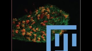
Another query- I have one nuclear (BrDu) marker and one cytosolic (DCX) marker, do you think I should add both binary and grayscale elements to my output image bcoz my DCX staining is not always extremely bright and background free and brdu is much better
You should try different settings and see what is best for quantification using plugin #2. But yes, with difficult staining sometimes using that (DCX) as grayscale output can be smart. I would try out using BRDU as binary, with "convert to outlines" option, and adding DCX as either binary or grayscale. Then during counting with plugin #2, you can try to auto-dected only "inside outlines".
Thank you. Please check out the article that came with this and search for the keyword stereological. We briefly discuss it. However, the counting too has option for marking cells in 3d space, so it is kindof possible. See the checkboxes near top right in that tool, for 2d/3d modes.


Thanks you for you science public service! Really amazing
Very cool tools, will try them soon! Thanks!
Thank you very much Anders, your work is amazing ^^
great sharing peeps.
Thank you indeed, Anders. Weldone!
Thank you! Very interesting and useful!!!
Hello, do you have macro that does this ? Thank you very much for your amazing tuto !!
Dr. Lunde, I have a query. How do I precisely determine the correct threshold for my images? Do I need to keep the value constant for the entire experiment?
Good question. There are different opinions about this, and it depends on your scientific question and your image material. It is best if the value is constant for the entire experiment. This generally requires constant microscope settings. You can manually find the correct threshold by making sure that all of the background is removed. Or you can try an automatic threshold procedure, but they can give a bit unexpected results for some images, so I would be careful with that. You could also read this and other sources, and discuss with supervisors/colleagues: imagej.net/imaging/principles#considerations-during-image-segmentation-binarization
@@anlu2 thank you for response! Is there any way i can contact you? Will it be possible for you to guide me in the analysis of my images. I have highly pixelated confocal large images taken at 60x.. it will be a great help in my work that is stuck for almost a year. Thanks..
@@ud1819 lunde.anders@gmail.com. please send at least 1 example image, and tell me what you need to count.
@@anlu2 thank you Dr. Lunde. I will email you shortly with the required details and images.
Another query- I have one nuclear (BrDu) marker and one cytosolic (DCX) marker, do you think I should add both binary and grayscale elements to my output image bcoz my DCX staining is not always extremely bright and background free and brdu is much better
You should try different settings and see what is best for quantification using plugin #2. But yes, with difficult staining sometimes using that (DCX) as grayscale output can be smart. I would try out using BRDU as binary, with "convert to outlines" option, and adding DCX as either binary or grayscale. Then during counting with plugin #2, you can try to auto-dected only "inside outlines".
Hi, many thanks for this amazing tool, and congratulations for your paper. Do you think this could be compatible with stereological counting?
Thank you. Please check out the article that came with this and search for the keyword stereological. We briefly discuss it. However, the counting too has option for marking cells in 3d space, so it is kindof possible. See the checkboxes near top right in that tool, for 2d/3d modes.
@@anlu2 Many thanks.