one thing i want to say dat..u r far far better than those pg coaching teachers...if u r going to teach us d whole optha..m damn sure u r going to hit other teachers out of the market...excellent job...
Excellent!! Love how you've explained things in logical and practical frameworks that help understanding, and not just route learning- thank you so much!
Thanks a million dear Pranesh Bhai! You are excellent teacher and are doing a wonderful job. I wish you a very good health and everlasting happiness. Please continue teaching us.
On 34:40 you said that in macula the ration of gc to cone are 1:1 , But earlier in this video you noticed that there are 6.5 million of cones , so how can it be 1:1 ? Thank you in advance for answering. And thank you a lot for this extraordinary video
Brilliant!! Really liked the video. But i honestly preferred the white board video.But its actually fine, whatever is easier for you.At the end, the information is what matters
Sir great teaching sir...sir please please please please please please please please make some time out and make videos on 2 topics... Retina and squint
very nice lecture sir, Thank you.. but i have a doubt ..in Langsman embryology the time of choroidal fissure closer(cloboma)-- is during 7th wk.. and you mention it during sixth wk .. in mcq what wl be correct ans?
Ayesha Iqbal I want squint lecture to be complete and comprehensive ... so gimme some time. shall give u guys somethin worth watchin. till then watch Tim Root s video on strabismus on TH-cam. read Parsons on squint.
Bro, fovea is considered as the thinnest part - 0.10 mm compared to ora - 0.12 mm. Not much of a difference but still, for ur exams keep fovea/ foveola (central floor of fovea) as 1st choice. Ref. Kanski pg 580
DR PRANESH THE inner layer of optic cup you are drawing is the invaginated layer of cup itself and no other innner layer . your concepts are not correct i feel
ajit thakur ...True 😊 I felt the same too. I wanted to experiment with this one where I cud draw and display pics at the same time... But anyday white/blackboard teaching beats any technology out there. Thanks for the feedback


one thing i want to say dat..u r far far better than those pg coaching teachers...if u r going to teach us d whole optha..m damn sure u r going to hit other teachers out of the market...excellent job...
Excellent!! Love how you've explained things in logical and practical frameworks that help understanding, and not just route learning- thank you so much!
You are really good at ophthalmology and ur way making students understand the concept is simply awsome. More strength and power to u sir
Thanks a million dear Pranesh Bhai! You are excellent teacher and are doing a wonderful job. I wish you a very good health and everlasting happiness. Please continue teaching us.
I can't emphasize enough on how thankful I am for ur lectures..Sine joining residency I have been watching ur videos..pls continue making mor videos.
sir took saving lives a bit too seriously....THANKYOU SIR..the best ophthal lecture Ive ever watched
Very good communication and good skill of explaining keep it up and help to required person through your vedio sir
On 34:40 you said that in macula the ration of gc to cone are 1:1 ,
But earlier in this video you noticed that there are 6.5 million of cones , so how can it be 1:1 ?
Thank you in advance for answering.
And thank you a lot for this extraordinary video
This helped me a lot !.... thank you very much, regards from Saudi Arabia.
Thnqqq so much your videos is awsm so easly you define topics. Your lectures help me a lot
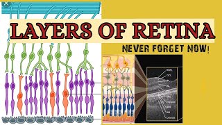
Really this video helping me to understand the anatomy of retina very well.thank you so much sir
Brilliant!! Really liked the video. But i honestly preferred the white board video.But its actually fine, whatever is easier for you.At the end, the information is what matters
excellent !!! keep it up very high yielding
Thank you for your quality time..
Nicely explained..
Means a lot to come from u, sir ! Thanks 🙏🏻
Pranesh
When u find time, just send a "hello" to my Whatsapp : 7076674212
Sure sir ! 😊😊
THANK YOU SIR
REALLY IT HELPED ME IN UNDERSTANDING THE RETINA
Excellent explanation! Thank you sir .
Awesome lecture sir
very nice ... continue your teaching Pranesh ..
Prem Kumar thank u thalaiva :)
Really well explained
Sir... Please add a video on Neurophthalmology.... And Thnkss for making my concepts clear on retina 😇
its good u started uploading videos again....can u plz make a video on strabismus? got an exam in optho in september...thanks...
Very well explained sir, indeed it helped me a lot
Brilliant work. Can you upload more ophthalmology video's as well. These video's helps us students to grasp a clear concept.
Sir great teaching sir...sir please please please please please please please please make some time out and make videos on 2 topics... Retina and squint
It's just excellent,very well explained each detail sir...
Very nice video sir
Wonderful explanation of a very difficult subject
I prefer this better than whiteboard..excellent lecture sir..i hav exms nxt mnth..could u pls make a topic on diseases of retina sir..
Beautifully explained
excellent! please upload more! From Bangladesh!
fantastic lecture many thanks sir
Excellent lecture thank u .......sir pls do strabismus topic for pg aspirants...
Helpful
OUTSTANDING LECTURE
thank you soo much Sir you are inspiration you help me understand retina please can you do more video thank you again good luck
Grt job👍
More vedios plz...
well explained Friend. thank you
Amazing video.. Thank you sir.
It is very understandable ...thank s sir
Comprehensive video. Wish you would do same for Uveal Tract and Sceral/Cornea. Thanks so much for this
Really liked it
what's papelledema
THANK YOU SO MUCH
Thankyou so much
Plzz make more ophtha videos for ug medical students !!
tq so much !
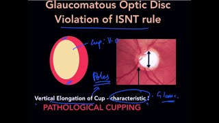
Optic cup and optic disc ratio, what if it’s .0.06=60% is that normal range, when does it become not normal range?
what book are the images from the "gross anatomy of the retina" from? Thanks, you really explain this topic well.
Clinical anatomy of eye - Lee Ann Remington
very nice lecture sir, Thank you.. but i have a doubt ..in Langsman embryology the time of choroidal fissure closer(cloboma)-- is during 7th wk.. and you mention it during sixth wk .. in mcq what wl be correct ans?
7 wks 😊
Sir pls make a video on glaucoma.... Plsss..... 🙏🙏
Super sir
Plz plz make video on conjunctiva squint and lens
Sir I cannot download the pdf
Can you upload a video on retinitis
part 2 angle of ant chamber?
so can plz upload a lecture on strabismus?
Ayesha Iqbal I want squint lecture to be complete and comprehensive ... so gimme some time. shall give u guys somethin worth watchin. till then watch Tim Root s video on strabismus on TH-cam. read Parsons on squint.
@@pranesh you are a good person👍🏽
Sir can you please upload extra edge PDF
Thank you sir
From where can i get the extra edge pdf key points on this topic
The link is not supporting plz provide the valid link
yea i guess file access expired. shall upload a newer one soon n let u know.
@@pranesh yes do mention sir & tag me so that I'll come to know thanku
Sir can u upload new link of pdf file as u said u will upload it soon.. Please kindly provide it
@@bhagyashreegawde2942 drive.google.com/file/d/1s3irBFv-FDSICUx1kuPwm96v7riXcwth/view?usp=sharing
Thinnest portion isnt it ora serrata? I’ve seen everyone saying ora serrata now I’m confused
It's foveola
Sir isnt ora serrata the thinnest part of retina ??
Bro, fovea is considered as the thinnest part - 0.10 mm compared to ora - 0.12 mm. Not much of a difference but still, for ur exams keep fovea/ foveola (central floor of fovea) as 1st choice. Ref. Kanski pg 580
Thank u sir 😃
@@pranesh thanks. I would definitely read Kanski
3:59 - you have made wrong diagram
DR PRANESH THE inner layer of optic cup you are drawing is the invaginated layer of cup itself and no other innner layer . your concepts are not correct i feel
I wana know if this guy can make me a slushy
If u provides whole opth lectures for pg aspirant with some money let me know.
your previous white board teaching was far better than this power point teaching....
ajit thakur ...True 😊 I felt the same too. I wanted to experiment with this one where I cud draw and display pics at the same time... But anyday white/blackboard teaching beats any technology out there. Thanks for the feedback
ajit thakur ,u maybe right but white board teaching is almost vanished fart in the wind. It's new era of teaching.
the concept of retinal layer is not even clear for you
Thank u so so much