MR neurography is a very useful addition to the usual MRI c spine. The fat in the nerves is emphasised and lesions of the nerves are much clearer. That having been said this was an excellent tutorial. Thank you
This is awesome. I had a collapsed cspine and have the images in my phone. I paused the video to go look at my images after each segment and not sure if the doc is an instructor but he should be. I followed along and as a nurse reading images has always been challenging until now. Grateful for the explanation and new knowledge!
Hello hope ur doing well Do u have torn muscle and ligament in ur neck . Its been 2 years . But its getting worse and worse. My mother is suffering with this excruciating pain . Pain radiates both her hands , tingling in neck and numbness in finger . Do u also have these symptoms. Plz reply me if you found any help
Fails to highlight vertebral facets joints and UV joints (Uncovertebral joints). These joints can enlarge or become less flexible and thus narrow the foramen. The video correctly notes that a narrowed foramen can compress a nerve root, thus causing neck pain.
Hello, great video. I Just had my MRI results shown and told to me today and I was told I have a minor disk bulge on L5 however I noticed that my disks are very white above those points but that disk bulging along with my S1 are much much darker than the rest. I have very bad back pain and left leg tingling and pain occasionally but daily. Any information in your personal opinion would help me understand what may be happening. I was to nervous and a bit rushed so I didn't ask the questions I should have. DDD does seem to run in family on both sides However I am quite young.
Looks like cervical spinal cord MRI picture covers more than just the cervical section - also brain stem and upper back. Is it still just as informative and accurate for those areas as well? I mean the whole brain stem and upper back segment
Can you actually visualize the nerves and nerve roots themselves coming off the cord and measure how inflamed they are? Or can you only measure the size of the space between the foramina?
Hello, I just got my MRI and it says that I have a 3 mm l5-s1 circumstantial disc bulge with mild lower neural foraminal narrowing, central canal is normal. I am having pretty severe discomfort on my central lower back when sitting down or driving. Can anybody help to explain my situation?any help would be appreciated, since I have stopped working because of it. Thank you.
L5-S1 disc bulge is compressing the exiting nerve roots, this may be the cause of your pain. You need to see your doctor for advice and treatment, including instructions about the correct way to sit, not bending or carrying heavy weights etc.
@@alsawiyusuf3192 hello.it appears that it might be a facade joint issue. I am going to see a pain management specialist,it looks I might need an injection which I'm trying to avoid. hopefully the pain specialist will be able to help. Thank u for the help, appreciate it.


![ภาพนี้ก็ฮาเหมือนกันนะเนี้ย #1 SS8 [ พากย์นรก MEME.EXE ] | easy boy](http://i.ytimg.com/vi/Ytc50m91QK8/mqdefault.jpg)


MR neurography is a very useful addition to the usual MRI c spine. The fat in the nerves is emphasised and lesions of the nerves are much clearer.
That having been said this was an excellent tutorial. Thank you
thank you for your wonderful lectures.
Thank you. I believe I can read my own MRI now.!
BEST ever!!!!! thank you .
Excellent review of mri
Thank you
Thank you from a new surgical neuro Nurse Practitioner
Thanks. That was an informative video.
Thank you! I can see it now.
Thank you this helps so much!!
This is awesome. I had a collapsed cspine and have the images in my phone. I paused the video to go look at my images after each segment and not sure if the doc is an instructor but he should be. I followed along and as a nurse reading images has always been challenging until now. Grateful for the explanation and new knowledge!
Hello hope ur doing well Do u have torn muscle and ligament in ur neck . Its been 2 years . But its getting worse and worse. My mother is suffering with this excruciating pain . Pain radiates both her hands , tingling in neck and numbness in finger . Do u also have these symptoms. Plz reply me if you found any help
So helpful thank you.
Thank you for taking the time to help 👍🏼👍🏼
THANK YOU!
I just got a new mri this week. I am waiting for the results.
It's a great lecture.
I'd like an X-RAY lecture, too.
Thanks!!😄
Short and sweet, nice!👍
Thanks a lot
Fails to highlight vertebral facets joints and UV joints (Uncovertebral joints). These joints can enlarge or become less flexible and thus narrow the foramen. The video correctly notes that a narrowed foramen can compress a nerve root, thus causing neck pain.
Great video! Thank you for sharing. 👍
Sir please make a video about neck capillary vassals
Hello, great video. I Just had my MRI results shown and told to me today and I was told I have a minor disk bulge on L5 however I noticed that my disks are very white above those points but that disk bulging along with my S1 are much much darker than the rest. I have very bad back pain and left leg tingling and pain occasionally but daily.
Any information in your personal opinion would help me understand what may be happening. I was to nervous and a bit rushed so I didn't ask the questions I should have.
DDD does seem to run in family on both sides However I am quite young.
awesome , thank you so much
Looks like cervical spinal cord MRI picture covers more than just the cervical section - also brain stem and upper back. Is it still just as informative and accurate for those areas as well? I mean the whole brain stem and upper back segment
Can you actually visualize the nerves and nerve roots themselves coming off the cord and measure how inflamed they are?
Or can you only measure the size of the space between the foramina?
How do you see the alar and transverse ligaments of c1?
Do all radiologists look at the cross sections like your doing.
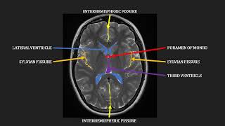
So the cervical MRI shows the base of the brain?
Hello, I just got my MRI and it says that I have a 3 mm l5-s1 circumstantial disc bulge with mild lower neural foraminal narrowing, central canal is normal. I am having pretty severe discomfort on my central lower back when sitting down or driving. Can anybody help to explain my situation?any help would be appreciated, since I have stopped working because of it. Thank you.
L5-S1 disc bulge is compressing the exiting nerve roots, this may be the cause of your pain. You need to see your doctor for advice and treatment, including instructions about the correct way to sit, not bending or carrying heavy weights etc.
@@alsawiyusuf3192 hello.it appears that it might be a facade joint issue. I am going to see a pain management specialist,it looks I might need an injection which I'm trying to avoid. hopefully the pain specialist will be able to help. Thank u for the help, appreciate it.
Easily explained
👌🏵️T2 : 03:02 :
🔯[縱軸][中心椎管Central canal]狹窄03:36~03:50
🔯 [側向][椎間孔IVF]狹窄
🌹👍 03:52 & 04:06~05:10在T2縱切影像系列的兩側查找IVF,正常應可見灰黑色圓形頸神經被一團佔據整個IVF的白色脂肪組織圍繞。❇️當某節椎孔狹窄時,原應為脂肪組織所在空間便因被前方椎間盤(呈像黑)膨出佔據而白色訊號消失。04:18
🔯再仔細看個別椎盤的橫切面05:11 05:22
Foramen椎孔5:39
🌹此處有椎盤向右側椎孔凸出 6:34
Yeah
No sound
🖒
Samajh me achche sebnahi aara please
Bhai hindi mein video banao na